Cuneiform fractures
Introduction
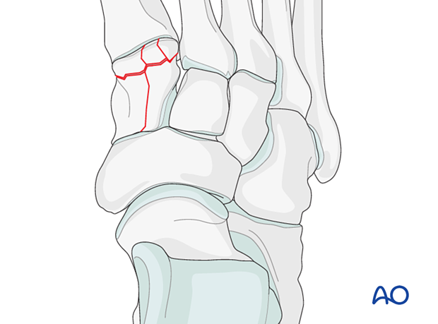
Cuneiform injuries are bound by the Lisfranc joint distally and the Chopart joint proximally. They may be isolated or, more commonly, involved in a Lisfranc injury.

Clinical presentation
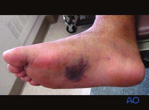
There is swelling, bruising, and point tenderness.
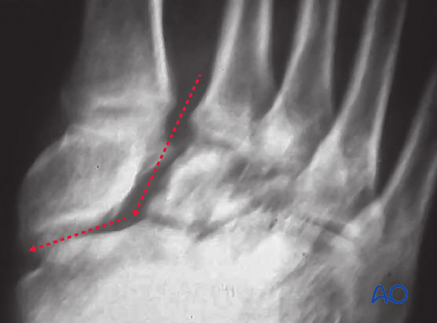
Complete intraarticular fractures may be associated with a shortening of the medial column.
In the multi-injured patient, foot fractures are often overlooked and are picked up on the secondary survey. In the unconscious patient, one must rely on a careful physical examination. Swelling, crepitus, or a deformity are suggested signs of underlying injury and should be followed up with appropriate x-rays.

Images
Plain x-rays will often show the injury pattern.
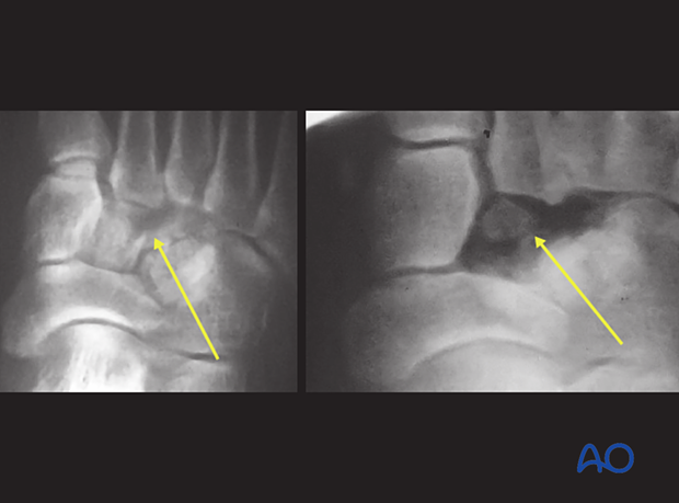
CT with sagittal and coronal reconstruction helps obtain a three-dimensional understanding of the injury. CT protocol should be thin cuts with significant overlap.

Mechanism of injury
These injuries may arise in athletics and be very subtle, or they may result from higher-energy injury and be more obvious. Their mechanism is similar to a Lisfranc midfoot injury, and they may indeed be part of Lisfranc fracture-dislocation.
The high-energy injury is often part of a polytrauma and is often associated with other injuries in the foot and other body parts.

Associated injuries
Isolated cuneiform injuries are rare, and they are often a part of a Lisfranc injury. A high index of suspicion is required to exclude a Lisfranc injury.


