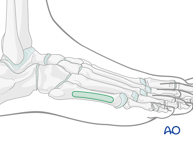
Safe zones for pin and K-wire insertion in the foot
1. Introduction
2. Medial safe zones
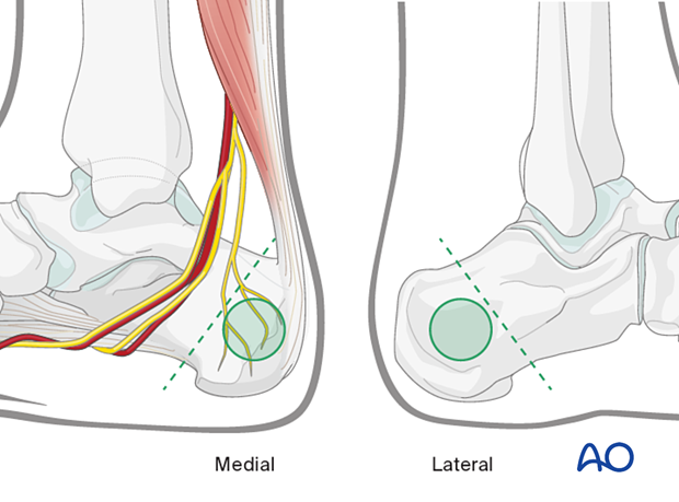
Calcaneum
The safe zone is behind the green dotted line shown in the figure.
If swelling permits, locate the posterior tibial pulse: if this is not palpable with certainty, examine the pulse of the uninjured leg and use its position as a guide to the probable path of the bundle at the injured side.
Pins inserted into the calcaneal tuberosity from medial to lateral minimize risk to the neurovascular structures behind the medial malleolus (posterior tibial artery, medial and lateral plantar nerves, and the medial calcaneal nerve).
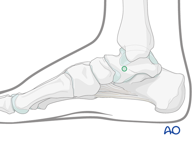
Talar neck
The insertion point is in the talar neck. The neck is found by palpation, and it is located midway between the navicular tubercle and the tip of the medial malleolus.
Care must be taken not to injure the articular surface of the talus.
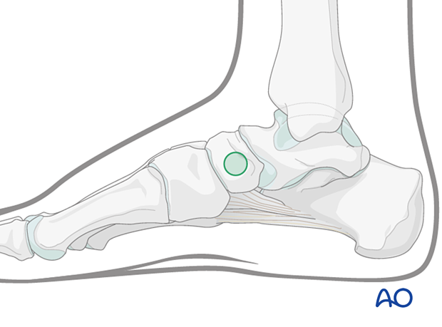
Navicular
A safe insertion point is located slightly distal to the navicular tubercle.
Care must be taken not to penetrate the concave articular surface of the navicular. Pin direction is medial to lateral.
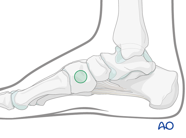
Cuneiform
If used, the insertion point is typically centrally at the medial surface.
Pin direction is medial to lateral.
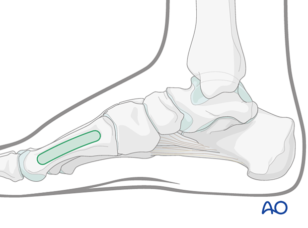
Medial aspect of the first metatarsal
The safe zone is located anywhere along the medial aspect of the first metatarsal.
Pin direction is medial to lateral.
3. Lateral safe zones
Cuboid
The cuboid is not used.
Calcaneum
The safe zone is behind the green dotted line shown in the figure.
If swelling permits, locate the posterior tibial pulse: if this is not palpable with certainty, examine the pulse of the uninjured leg and use its position as a guide to the probable path of the bundle at the injured side.
Pins inserted into the calcaneal tuberosity from medial to lateral minimize the risk to neurovascular structures behind the medial malleolus (posterior tibial artery, medial and lateral plantar nerves, and the medial calcaneal nerve).

Fifth metatarsal
The safe zone is located on the lateral aspect of the fifth metatarsal.
Pins are inserted from lateral to medial. If the 5th metatarsal is unstable, the pins can be inserted through the 5th and the 4th metatarsal for added support.
