Medial approach to the 1st metatarsal
1. Indications
The medial approach to the first metatarsal is used to access the entire length of the first metatarsal. The lateral aspect of the first metatarsal may be challenging to visualize through this approach.

2. Anatomy
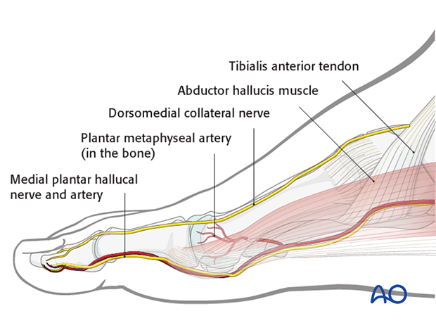
In the adult, the first metatarsal is supplied by an artery entering the first metatarsal head via the plantar distal metaphysis.
The courses of the dorsomedial collateral nerve (usually the superficial peroneal nerve) and the medial plantar hallucal nerve are illustrated.
The abductor hallucis tendon inserts into the medial capsule of the first metatarsophalangeal (MTP) joint.

3. Skin incision
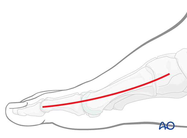
The skin is incised over the medial aspect of the metatarsal shaft, starting at the medial cuneiform and extending distally to the first metatarsophalangeal joint.

4. Exposure of the bone
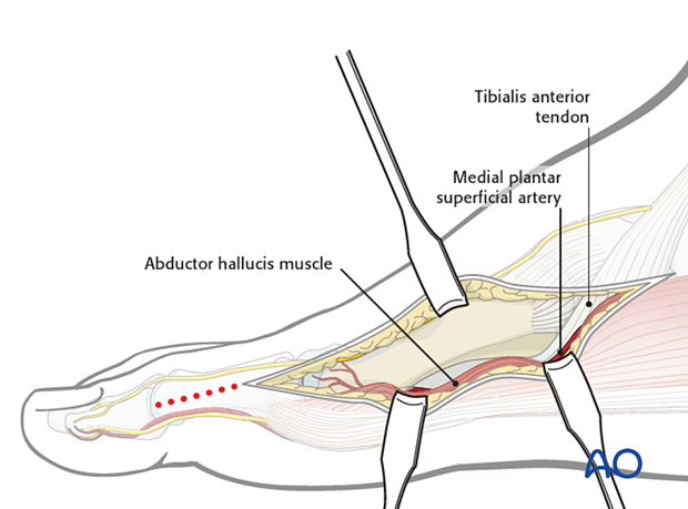
Find the dorsal edge margin of the abductor hallucis muscle and retract it plantarwards.
Free the distal part of the tibialis anterior tendon to gain access to the first tarsometatarsal joint.

5. Closure
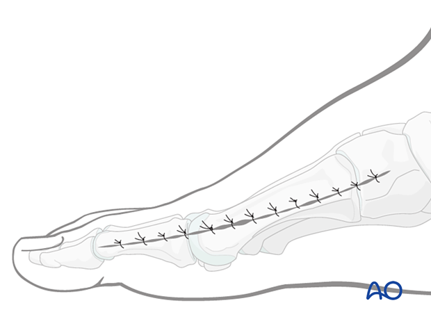
Perform a subcuticular closure with resorbable sutures.
The skin should be closed with appropriate sutures. Nylon sutures are typically used.


