Dorsal approach to multiple metatarsals
1. Indications
This approach provides access to multiple metatarsals.
2. Preservation of the vitality of the soft tissues and of the bone
A solitary incision over an individual metatarsal is safe.
If multiple metatarsals require access, individual incisions over each metatarsal should be avoided.
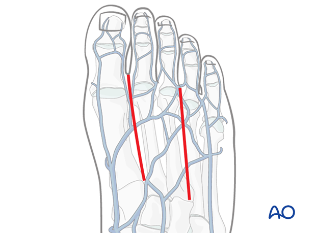
If several metatarsals are to be approached two incisions are used, one between the 1st and 2nd and/or one between the 3rd and 4th.

3. Anatomy
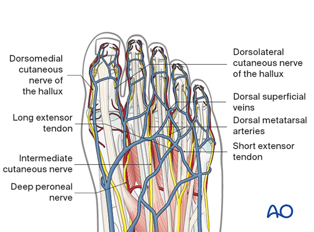
The veins are superficial and longitudinal veins should be preserved.
The long tendons lie superficial and medial, while the short tendons are deep and lateral.
Branches of the deep peroneal nerve which divides to provide the superficial digital innervation must be identified and protected in this approach.

4. Skin incision
Depending on the injury, the incision can be made between the 1st and 2nd metatarsal and/or the 3rd and 4th metatarsal.
Make a longitudinal incision extending it from the metatarsophalangeal to the tarsometatarsal joint.

5. Deep dissection
The approach to the dorsal aspect of the metatarsal is made in such a way as to protect the intermetatarsal nerves and the crossing superficial veins. The optimal approach goes inbetween the long and the short extensor tendon of the corresponding ray.
For this approach, no muscle should be incised. But, the muscle needs to be released off the bone.

6. Closure
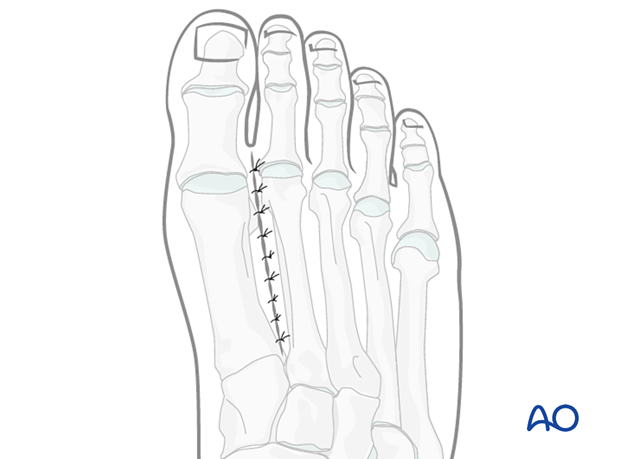
Close the joint capsule using resorbable sutures.
Repair the tendon sheath over the extensor hallucis tendon if possible.
Perform a subcuticular closure with resorbable sutures.
The skin should be closed with appropriate sutures. Nylon sutures are typically used.


