TMT (Lisfranc) injuries
Definition
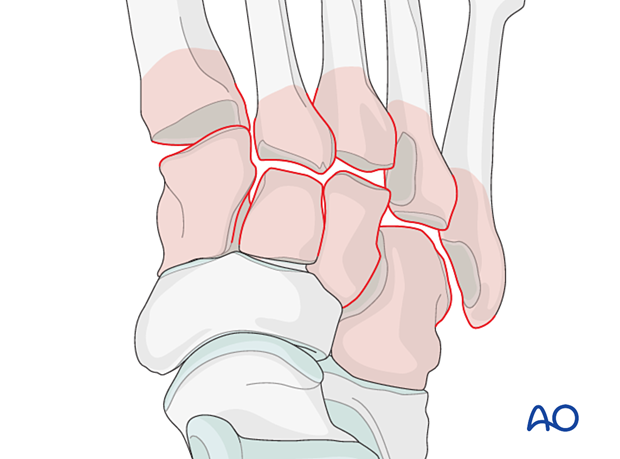
Lisfranc (tarsometatarsal) injuries cover a spectrum of injuries that may include any combination of:
- Injuries to the Lisfranc ligament, tarsometatarsal ligaments, and/or the intermetatarsal or intertarsal ligaments
- Fractures of the proximal metatarsals, cuboid, and/or cuneiform bones
Also, there may be associated injuries in both the forefoot, hindfoot, and ankle.
Clinical presentation
The typical clinical findings include:
- Dorsal midfoot swelling
- Pain upon palpation of the TMT area
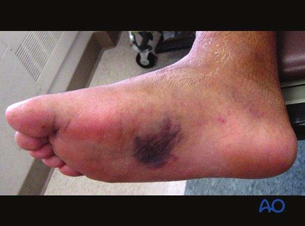
- Dorsal and/or plantar medial ecchymosis
- Instability
The pain is seemingly out of proportion to the injury. Unlike a routine ankle sprain, these injuries may be described as visceral.
Pitfalls
- Beware of a patient who was told in the Emergency Room that they had a "sprained foot".
- Since Lisfranc injuries may represent instability without frank displacement, ER x-rays (which are often non-weight bearing) may not show the extent of the injury.
- Subsequent weight-bearing x-rays in the office or clinic may show displacement/instability.
- The surgeon should have a low threshold for ordering a CT scan when in doubt.
- Remember that Lisfranc injuries frequently involve high energy, which dissipates through the soft tissues, and therefore they may be associated with a compartment syndrome.
Imaging
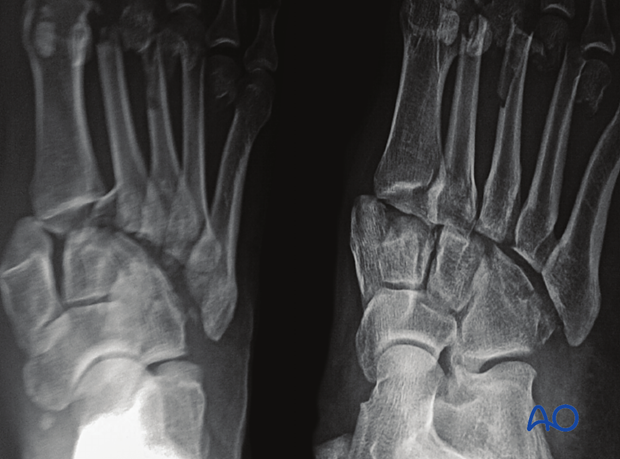
During the initial evaluation, up to 20% of Lisfranc fracture-dislocations are missed. Images must be recorded in AP, lateral, and 45° internal oblique views with associated weight-bearing views.
Characteristic x-ray features include:
- Misalignment of metatarsals with associated cuneiform cortices
- Bone chip at base of 2nd metatarsal
- > 3mm distance between the bases of 2nd and 3rd metatarsal
- Proximal metatarsal fractures
There may be additional X-ray features associated with injuries to:
- Cuneiform
- Cuboid
- Navicular
- The anterior calcaneal process (combined Chopart and Lisfranc injury)
- Ligament avulsions
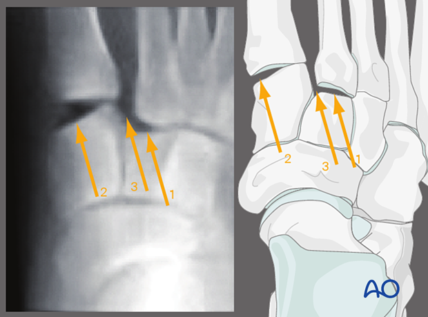
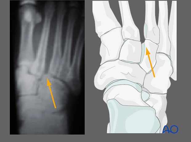
AP view
1. Lateral displacement of 2nd metatarsal on middle cuneiform
2. 1st TMT joint disruption
3. The gap between 1st and 2nd metatarsal and middle and medial cuneiform
45° internal oblique view
Lateral displacement of 4th metatarsal on the cuboid.

Lateral displacement of 3rd metatarsal on the lateral cuneiform.
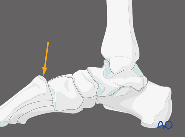
Lateral view
The dorsal cortex of the metatarsals should be even with the dorsal cortex of the cuneiforms. This image shows a dorsal displacement of the metatarsal bases relative to the cuneiforms.
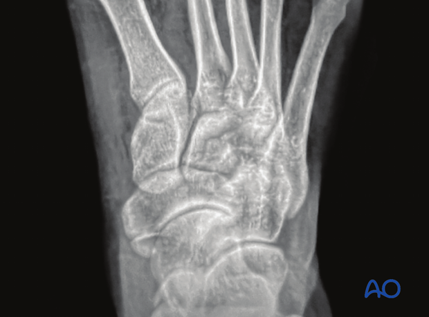
Weight-bearing views
If you suspect an occult injury, insist on a weight-bearing film within the patient's tolerance.
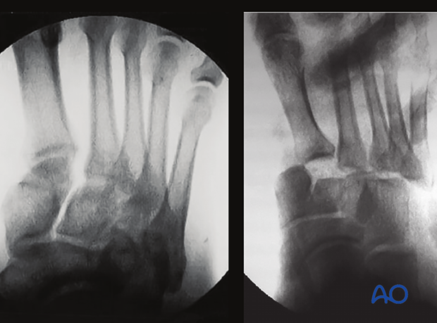
Stress X-rays
The intraoperative stress test can uncover instability that may not be apparent in a static x-ray. Stress testing is painful and is typically done intraoperatively.
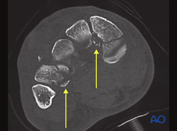
CT
CT scans and a 3D reconstruction can be helpful to define the injury further.
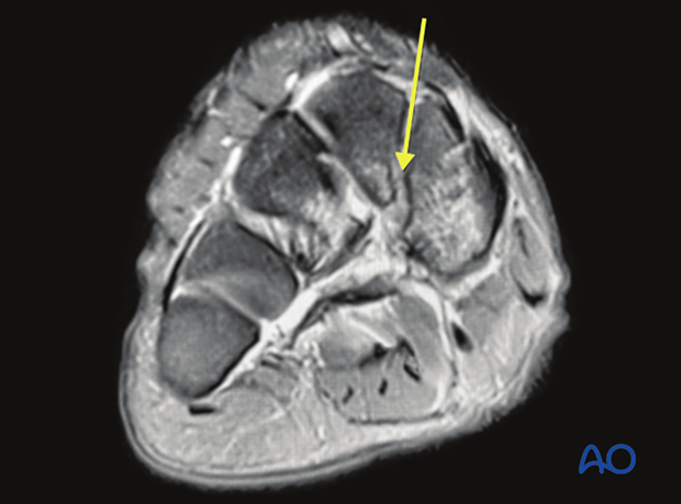
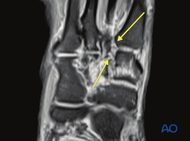
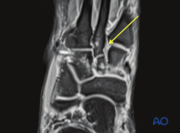
MRI
MRI may be indicated for the identification of pure ligamentous injuries.
Mechanism of injury
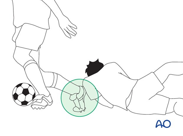
Direct or indirect forces may cause Lisfranc injuries.
Direct forces include a crush injury (MVA or industrial) or a direct blow. These may be combined with soft-tissue injury and present as open fractures.
An indirect mechanism is more common than a direct one. Indirect injuries result from an axial load to a plantarflexed foot, forced abduction of the forefoot, or both. They may occur during sports or stepping down a stair or sidewalk.
Associated injuries
There may be associated bony and ligamentous injuries in the forefoot, hindfoot, and ankle.


