Dynamic fixation of ruptured Lisfranc ligament
1. Introduction
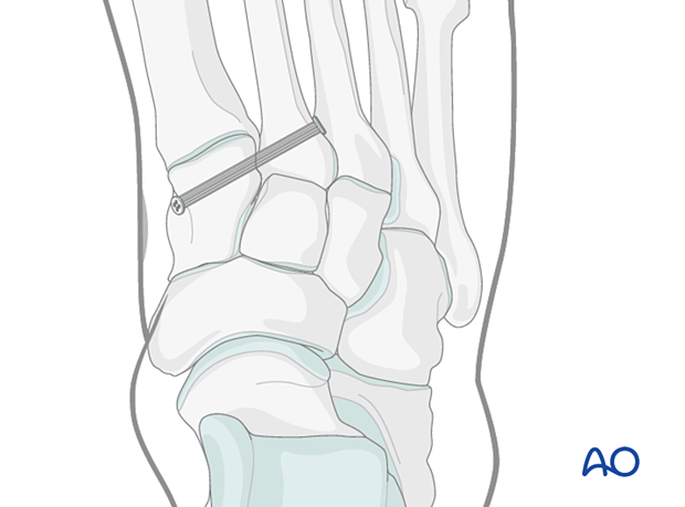
The principle of this procedure is to stabilize the anatomic relation between the base of the second metatarsal and the medial cuneiform using dynamic fixation. This fixation involves using suture tape, which is anchored against the bone using metal buttons medially and laterally.
A wide range of fixation devices exist for this procedure, and the technique for tightening the suture tape will vary.
2. Approach
This procedure is performed using both:
- A limited dorsomedial approach to expose the base of the second metatarsal, and
- A limited medial utility approach to expose the medial side of the medial cuneiform.
3. Reduction
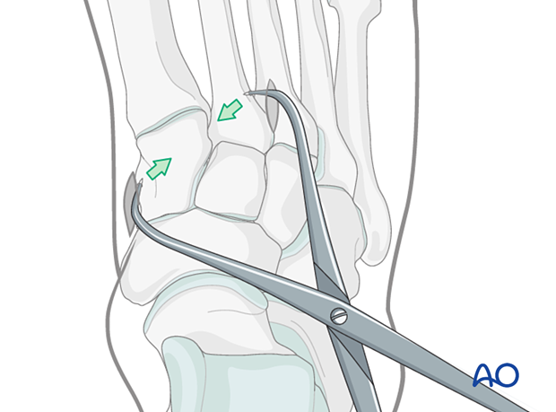
To facilitate the application of the reduction forceps, a K-wire may be used to create a recess laterally at the base of the second metatarsal and medially on the medial cuneiform.
Use pointed reduction forceps to reduce the second metatarsal base to the medial cuneiform. Be mindful to place the reduction forceps away from the placement of the planned position screw.
The reduction forceps can be used percutaneously and should be applied to provide compression in the vector of the Lisfranc ligament.
Verify the reduction visually and radiographically.
4. Fixation
Drilling
Insert a guidewire from the lateral aspect of the second metatarsal base. Under fluoroscopic guidance, direct it towards the medial cuneiform.
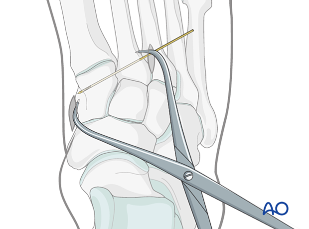
Drill over the guidewire using an appropriately sized cannulated drill bit.
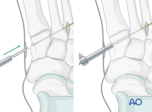
Insertion of the dynamic fixation device
Insert the dynamic fixation device and distal button from the cuneiform to the second metatarsal.
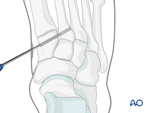
Tighten the dynamic fixation device to anchor the buttons both medially and laterally. Ensure that the buttons are securely anchored directly on the bone.
Verification of fixation
Verify reduction of the second metatarsal to the medial cuneiform and the button location under image intensification.


