Dislocation and fracture-dislocation of the metacarpophalangeal joint
Definition
Dislocations and fracture-dislocations of the metacarpophalangeal (MCP) joint often show dorsal dislocation.
The dislocations are classified by the AO/OTA as 70E1.2–5[5], where 2–5 indicates the ray of the injured MCP joint.

An MCP dislocation may be associated with soft-tissue injuries and fractures of the metacarpal and/or proximal phalanx.
The associated fractures may be a partial or complete articular fracture of the metacarpal head.

Imaging
X-ray
AP, oblique, and lateral views are required.
These AP and oblique x-rays show a case of a 2nd MCP joint dislocation with associated fracture of the metacarpal head.
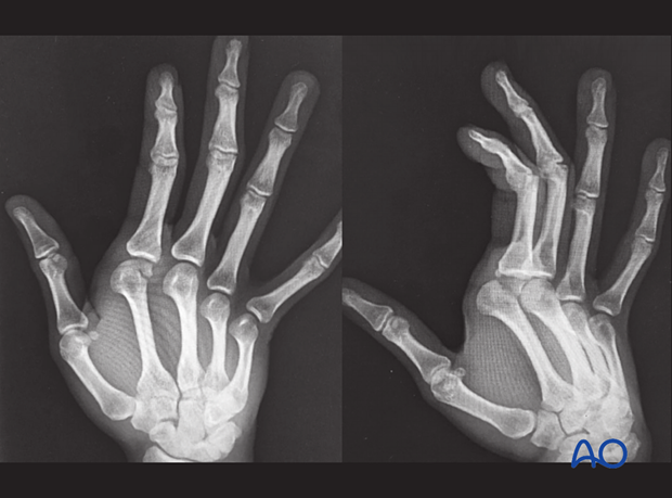
Dorsal dislocation is readily seen in the lateral or oblique view.
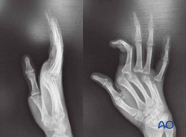
In this case, the AP x-ray shows dislocation of the 2nd, 3rd, and 5th MCP joint.
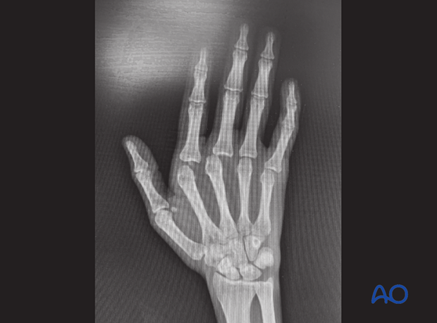
The extent of dislocation is better visible in the lateral and oblique x-rays.
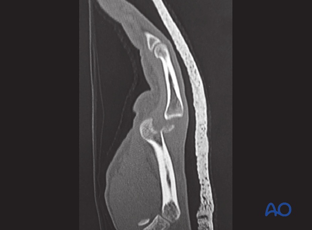
CT scan
CT scans help evaluate the size, shape, and pattern of an associated fracture.
This lateral CT image shows the MCP joint dislocation and associated metacarpal head fracture.
