Intramedullary screw fixation
1. General considerations
Introduction
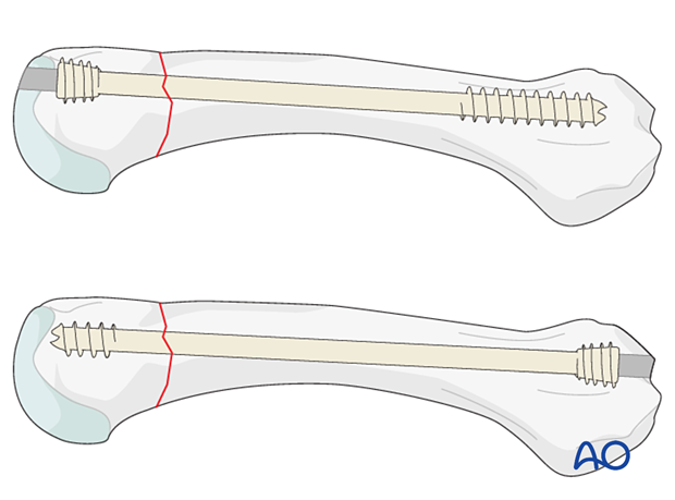
Transverse extraarticular neck fractures may be fixed with an intramedullary headless screw (3.0 or 4.0 mm), either ante- or retrogradely. The retrograde insertion needs thorough planning to avoid MCP joint problems.
The antegrade insertion needs at least 10 mm of bone stock in the distal fragment to get optimal fracture compression.
This screw acts like an intramedullary nail.
Prerequisites for intramedullary screw fixation
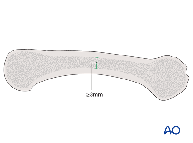
The diameter at the isthmus of the intramedullary canal should be at least 3 mm wide in AP and lateral views.
To allow for fracture compression, the length of the screw should be long enough so that the threads at the tip only engage in the far fragment.
To achieve optimal stabilization, the screw should be long enough and with a diameter filling the intramedullary canal.
2. Patient preparation
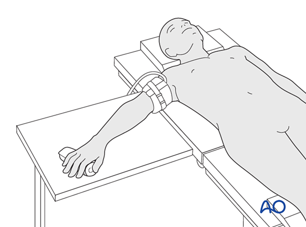
Place the patient supine with the arm on a radiolucent hand table.
3. Reduction
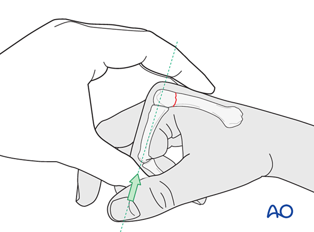
Reduce the fracture preliminarily by flexing the MCP and proximal interphalangeal (PIP) joints to 90° and using the proximal phalanx to push up the metacarpal head (Jahss maneuver).
Final reduction is performed with the guide wire and screw.
4. Retrograde screw insertion
Guide wire insertion
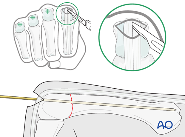
Hold the MCP joint in 90° flexion.
Incise the skin and extensor tendon and identify the dorsal part of the metacarpal head.
Insert the guide wire dorsally into the head in line with the intramedullary canal under image intensification until the tip reaches the base.
Confirm correct rotational alignment.
Screw insertion
Predrill the entry point and if needed the medullary canal. This creates a track for screw insertion and reduces the risk of deviation during screw insertion.
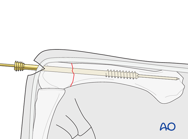
Select a screw with the appropriate diameter and length using the depth gauge provided. The thread of the tip should engage the isthmus distal to the fracture line.
Insert the screw over the guide wire until the head is fully entered into the metacarpal and the fracture is compressed.
Confirm reduction and correct screw placement with an image intensifier.
Confirm correct rotational alignment of the finger.
5. Antegrade screw insertion
Identifying the entry points
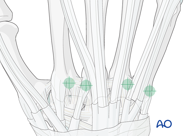
Identify the base of the metacarpal.
Incise the skin at the base of the metacarpal to expose its dorsal aspect.
Protect the extensor carpi radialis longus/brevis during the wire and screw insertion to the 2nd/3rd metacarpal.
Protect the extensor carpi ulnaris during the wire and screw insertion to the 5th metacarpal.
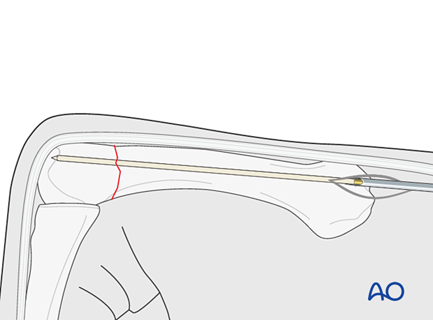
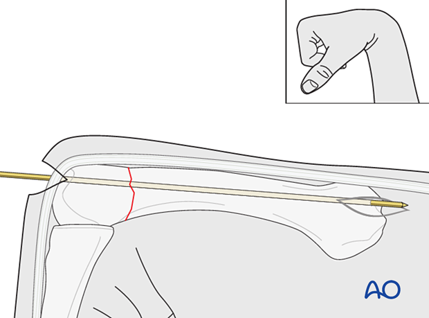
Guide wire insertion
Create an entry point with a drill to pass the guide wire into the bone.
Insert the guide wire dorsally into the base in line with the intramedullary canal under image intensification until the tip reaches the head.
Confirm correct rotational alignment.
Screw insertion
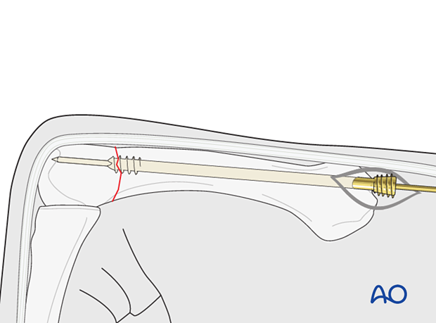
Predrill the entry point and if needed the medullary canal. This creates a track for screw insertion and reduces the risk of deviation during screw insertion.
Insert the selected screw over the guide wire until the head is fully entered into the metacarpal and the fracture is compressed.
Confirm reduction and correct screw placement with an image intensifier.
6. Final assessment
Confirm correct rotational alignment by clinical examination.
Image intensification may be used to confirm anatomical reduction and correct placement of implants in two views.
7. Aftercare
Postoperative phases
The aftercare can be divided into four phases of healing:
- Inflammatory phase (week 1–3)
- Early repair phase (week 4–6)
- Late repair and early tissue remodeling phase (week 7–12)
- Remodeling and reintegration phase (week 13 onwards)
Full details on each phase can be found here.
Postoperative treatment
If there is swelling, the hand is supported with a dorsal splint for a week. This would allow for finger movement and help with pain and edema control. The arm should be actively elevated to help reduce the swelling.
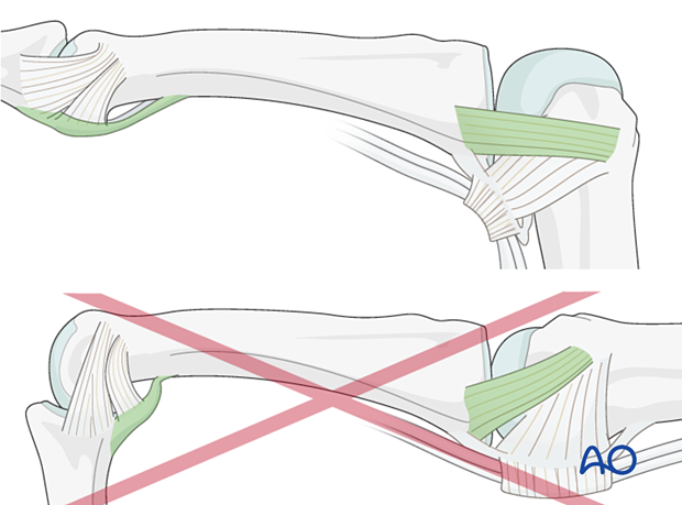
The hand should be splinted in an intrinsic plus (Edinburgh) position:
- Neutral wrist position or up to 15° extension
- Metacarpophalangeal (MCP) joint in 90° flexion
- Proximal interphalangeal (PIP) joint in extension
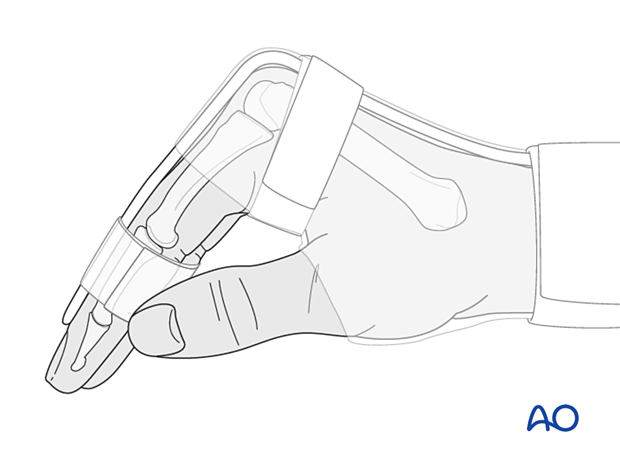
The reason for splinting the MCP joint in flexion is to maintain its collateral ligament at maximal length, avoiding scar contraction.
PIP joint extension in this position also maintains the length of the volar plate.
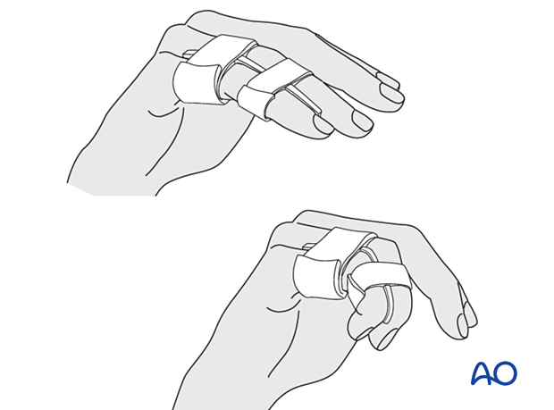
After subsided swelling, protect the digit with buddy strapping to a neighboring finger to neutralize lateral forces on the finger.
Functional exercises
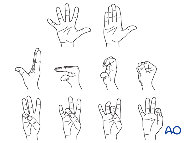
To prevent joint stiffness, the patient should be instructed to begin active motion (flexion and extension) immediately after surgery.
Follow-up
See the patient after 5 and 10 days of surgery.
Implant removal
Intramedullary screws should not be removed as this may result in cartilage damage.
8. Case
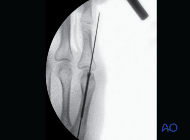
Intraoperative view of a reduced neck fracture of the 5th metacarpal with the guide wire and the cannulated intramedullary screw inserted through the metacarpal head
