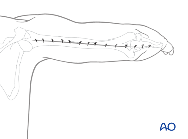
Posterior triceps-split approach to the humeral shaft
1. Introduction
This approach is most commonly used for fractures involving the distal half of the humerus. However, it can be extended for more proximal fractures once the radial nerve has been identified.
2. Skin incision
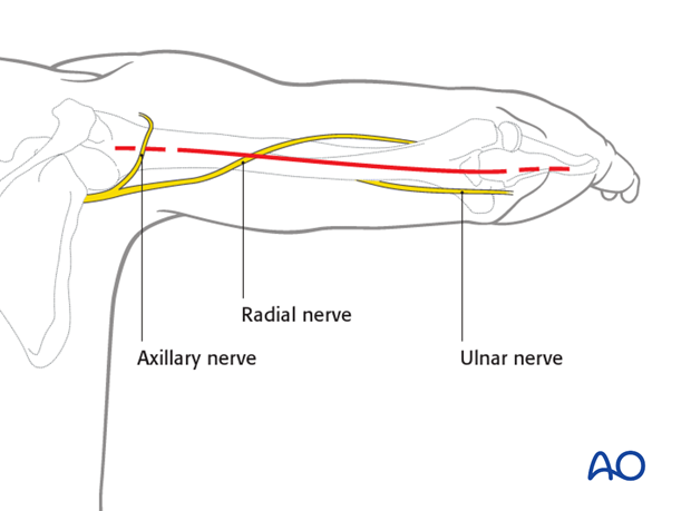
The complete incision is illustrated. Depending on the fracture and its location a smaller section might be used.
Incise the skin, beginning at the tip of the olecranon.
The incision runs proximally in a straight line along the posterior midline of the arm. It crosses the radial nerve in the mid-humeral region and the axillary nerve proximally.
3. Superficial dissection
Incise the deep fascia in line with the skin incision.
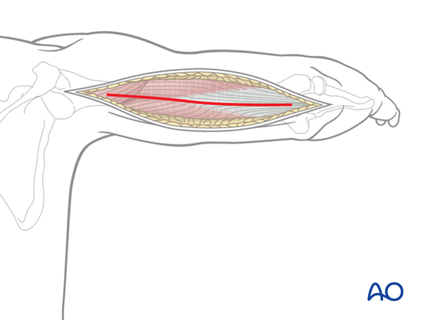
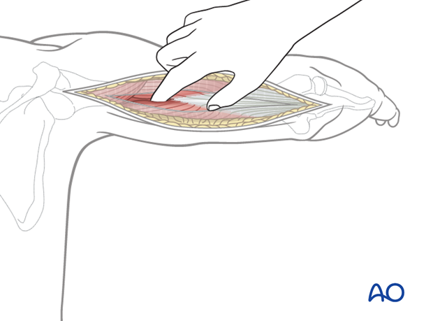
By palpation with a finger, identify the interval between the lateral and long heads of the triceps. The opening of this interval will be developed from proximal to distal, remembering that the radial nerve lies beneath the triceps as it crosses the humerus.
4. Deep dissection
Then develop the proximal interval between the two heads by dissection, retracting the lateral head laterally and the long head medially.
Within the spiral groove, identify the radial nerve and the accompanying profunda brachii artery.
Distally, split the common triceps tendon, along the line of the skin incision, by sharp dissection.
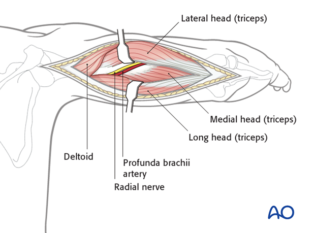
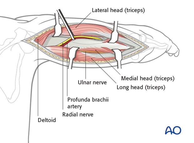
Release the lateral head of the triceps from the humerus proximally, and incise it distally, in line with the humeral shaft. Release the muscle from the bone only as much as needed and protect the ulnar nerve medially. The ulnar nerve is constrained at the point where it passes distally through the medial intermuscular septum, from the anterior to the posterior compartment of the arm.
Release the medial head of the triceps proximally until the axillary nerve is found.
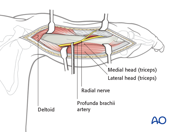
Now the posterior humerus, crossed by radial nerve and its accompanying vessels, lies exposed from the axillary nerve and posterior circumflex humeral artery proximally to the capitellum distally.
5. Extending the approach
Distal extension
Splitting the triceps tendon limits distal exposure, which can be improved by approaching the humerus from the lateral side of this muscle. For more details see the distal posterior approach.
Proximal extension
Limited proximal extension, even beyond the axillary nerve, is possible with careful mobilization and retraction of both radial and axillary nerves and their accompanying vessels.
6. Wound closure
Close the wound in layers in a standard manner.
