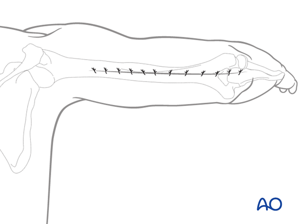
Posterior triceps-sparing approach (triceps-on) to the humeral shaft
1. Introduction
For midshaft and distal shaft fractures, the posterior approach may be extended distally, leaving the triceps insertion intact. This provides adequate exposure for reduction and fixation.
The triceps is elevated off the posterior humerus, but its insertion is not disturbed.
This retains the musculotendinous integrity of the triceps and allows more rapid postoperative rehabilitation.
2. Skin incision
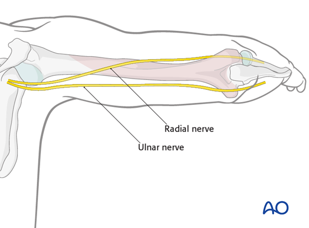
The complete incision is illustrated. Depending on the fracture and its location a smaller section might be used.
Incise the skin, beginning at the tip of the olecranon.
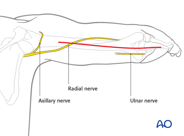
The incision runs proximally in a straight line from the olecranon along the posterior midline of the arm. It crosses the radial nerve in the mid-humeral region and the axillary nerve proximally.
3. Superficial dissection
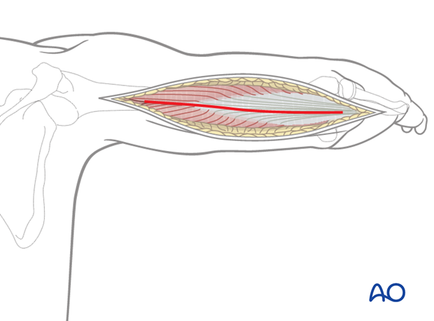
Incise the deep fascia in line with the skin incision.
4. Ulnar window
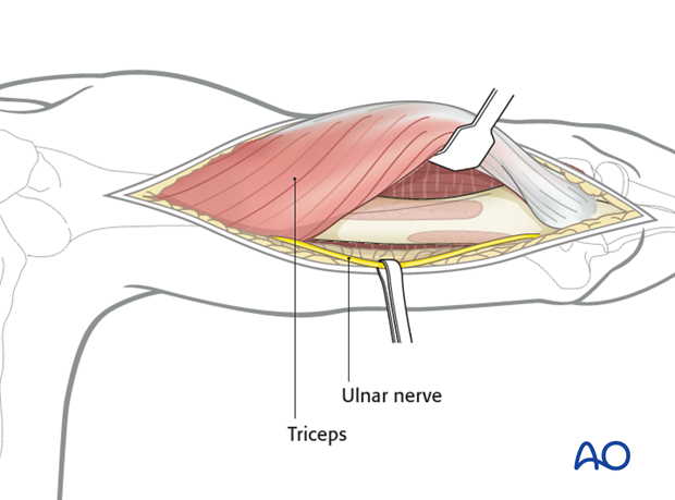
As a first step, identify and mobilize the ulnar nerve. It may be helpful to protect it with a vessel loop.
Proximally, follow the ulnar nerve along its course on the medial intermuscular septum.
Note: Take care that the protecting vessel loop does not injure the ulnar nerve by uncontrolled traction. Do not use heavy clamps to secure the loop.
Next, mobilize the triceps muscle and retract it laterally. This may be achieved by bluntly dissecting the medial head of the triceps from the posterior aspect of the humerus.
Depending on the fracture location the exposure may need to be extended distally.
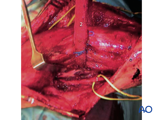
In the case shown here, going up into the diaphysis, the ulnar nerve was identified and held with a vessel loop (1).
The entire triceps muscle is isolated with a gauze wrap (2).
5. Radial window
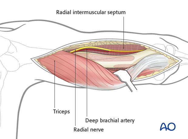
Split the triceps fascia and mobilize the lateral head of the triceps from the lateral intermuscular septum and humerus towards the ulnar side.
If necessary dissect remaining muscle fibers, still attached to the posterior aspect of the humerus, from the lateral side. This ends up in a liberated muscle complex containing the long head, lateral head and the medial head of the triceps. This permits the whole triceps muscle to be moved towards either the lateral or medial side, to provide access to the humerus (“triceps flip”).
The radial nerve can be detected at its penetration through the intermuscular septum and followed upwards in the radial groove.
6. Wound closure
Close the wound in layers in a standard manner.
