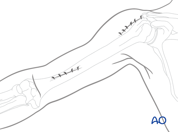
Limited approaches for anterior MIO of the humeral shaft
1. Introduction
The minimally invasive osteosynthesis approach allows for subcutaneous plate insertion through a tunnel. Typically, the plate will be introduced through the distal portal as shown below.
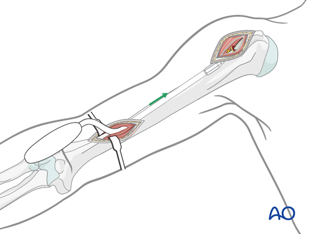
2. Distal portal
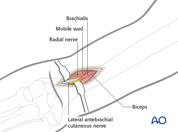
Distal access is gained through the brachialis muscle, splitting apart its medial and lateral portions for approximately 5 cm to reveal the anterior humeral surface. Distally the lateral antebrachial cutaneous nerve lies between biceps and brachialis. It should be identified and protected by medial retraction. The radial nerve lies laterally, protected by the lateral portion of brachialis.
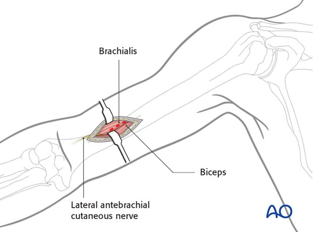
Brachialis has been identified deep to the biceps. Its fibers have been split longitudinally, providing extraperiosteal access to the anterior distal humeral shaft.
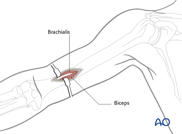
For more distal fractures, purchase on the lateral humeral column can be gained through a slightly more distal incision beginning at the joint crease and extending 5-6 cm proximally. Develop the interval between biceps and brachialis medially and the “mobile wad” (brachioradialis, extensor carpi radialis longus and brevis) laterally. The radial nerve must be protected here. Retract or incise the brachialis, as needed, to gain extraperiosteal access to the anterolateral aspect of the lateral column of the distal humerus.
3. Proximal portal (anterior deltoid split)
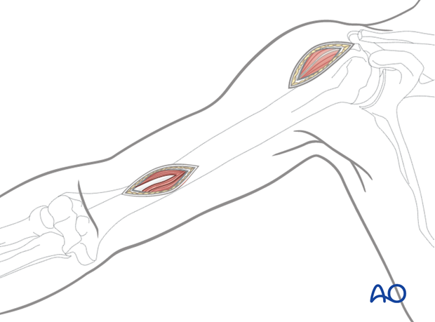
The fracture morphology determines the length of the plate required, and the site for proximal fixation. This dictates the site of the proximal portal.
For a more proximal fixation, expose the anterior deltoid raphe through an incision made distally from the anterolateral acromial tip.
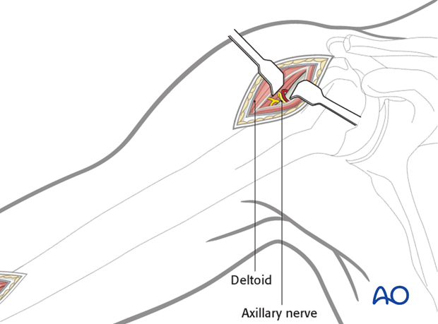
Carefully dissect through the raphe, finding and protecting the axillary nerve and its accompanying vessels. The proximal humerus lies beneath these. Mobilize this neurovascular bundle for extraperiosteal access to the proximal humerus.
4. Proximal portal (deltobicipital)
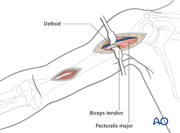
For shorter plate application in more distal fractures the humerus may be approached through the interval between the deltoid and biceps. The cephalic vein lies in this interval. Identify the vein and protect it while dissecting through the interval.
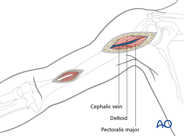
Dissect bluntly to the periosteal surface. Develop an area on the periosteal surface approximately 5 cm long. It is possible to release the anterior part of the deltoid insertion, if necessary, as this area of deltoid insertion is both long and broad.
5. Creating the tunnel
Create the extraperiosteal tunnel under brachialis with an instrument passed from the distal to the proximal incision.

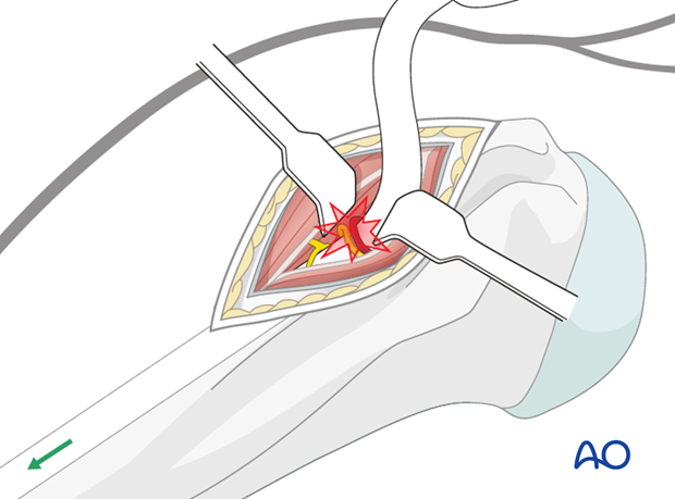
6. Pitfall: nerve damage
Take care not to injure the axillary nerve and its accompanying vessels especially when working from the proximal portal.
7. Wound closure
Close the incisions in layers in a standard manner.
