Complete articular fracture of the proximal end segment
Definition
Complete articular fractures of the proximal phalangeal base are classified according to AO/OTA as 78.2–5.1.1C, where 2–5 indicates which finger is injured.
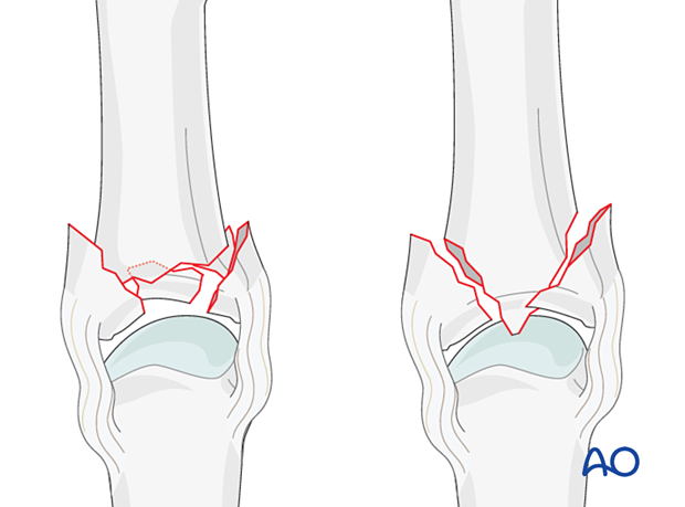
Typically, complete articular fractures occur in two configurations:
- Centrally impacted, comminuted fracture (multifragmentary)
- T- or Y-type fracture (simple)
When vertical compression forces, created by axial load, are applied to the finger, comminuted, intraarticular compression fractures may result. These fractures are very unstable.

Imaging
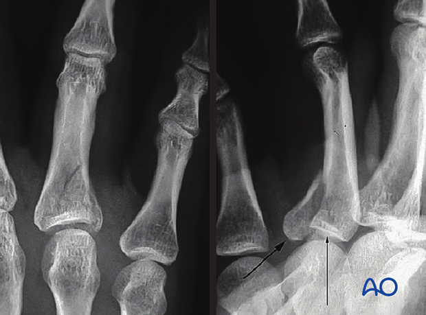
AP and oblique x-ray showing a complete articular fracture with impaction of the articular surface.

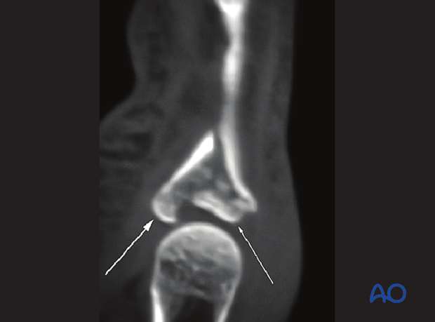
A CT image may provide information about the extent of impaction.
In both images, the left arrow indicates the volar cortical rim of the proximal end segment displaced in a volar direction. The right arrow indicates the articular surface tilted in a volar direction. The AP x-ray image shows that the articular surface is tilted in an ulnar direction. The combination of volar and ulnar tilts suggests that rotation of the fragment has occurred. This is an example of a ‘split-depression fracture’ of the load-bearing surface of a joint.


