Patient assessment
1. Introduction
Hand surgery involves a multi-specialty approach to the assessment, treatment, and aftercare of hand trauma. Therefore, the specialized teams should be involved early on according to the specific injury requirements.
A diagnosis is made based on the history, the mechanism of the trauma, clinical examination, and by x-rays in two planes.
The outcome of injuries of the fingers may be assessed by three criteria:
- Functional return
- Clinical assessment
- Radiological healing
Recommendations for the definition of extraarticular phalangeal fracture displacement vary. In the following, the definitions of the important relevant displacement are:
- No malrotation is accepted
- >2 mm of translation or axial displacement
- >10° of angular displacement
Reference: Freeland AE, Geissler WB, Weiss APC. Surgical treatment of common displaced and unstable fractures of the hand. Instr Course Lect. 2002:51:185–201.
2. Clinical assessment
Check for:
- Swelling
- Open wound
- Loss of sensation in the fingers (indicating nerve injuries)
- Perfusion (reperfusion of more than 2 seconds may indicate vascular injuries)
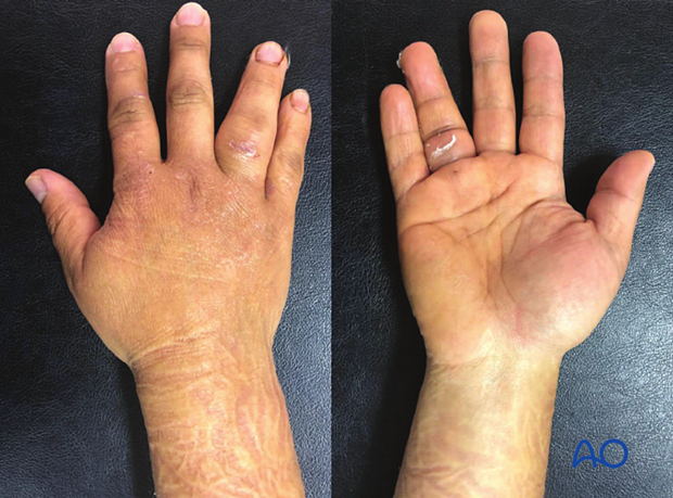
General assessment of hand and wrist
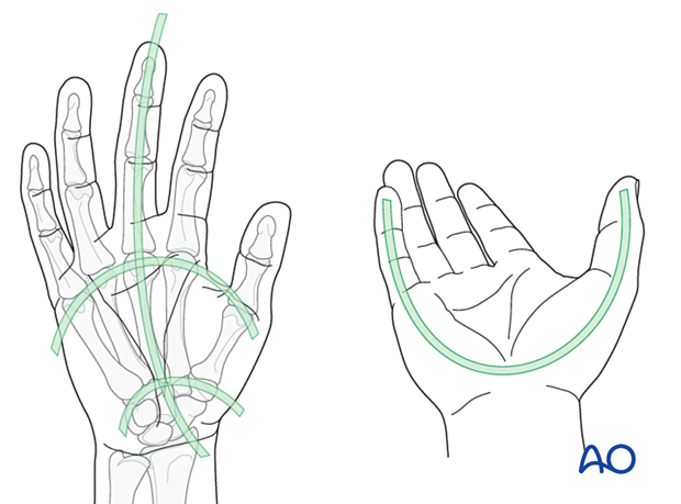
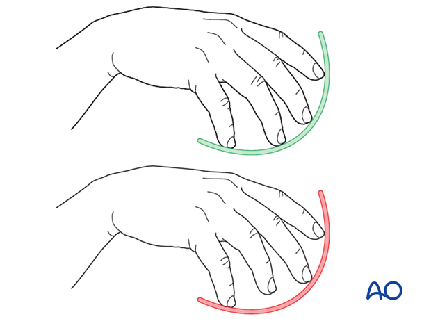
Check for anatomical architecture (position and relation) of the whole hand:
- Longitudinal arc
- Metacarpal arc
- Oblique arcs (with thumb in opposition)
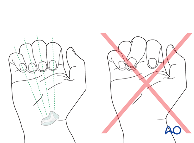
The tips of the flexed fingers should point to the scaphoid. Check for deviating or overlapping fingers. This will indicate malrotation.
Assessment of the proximal phalanx
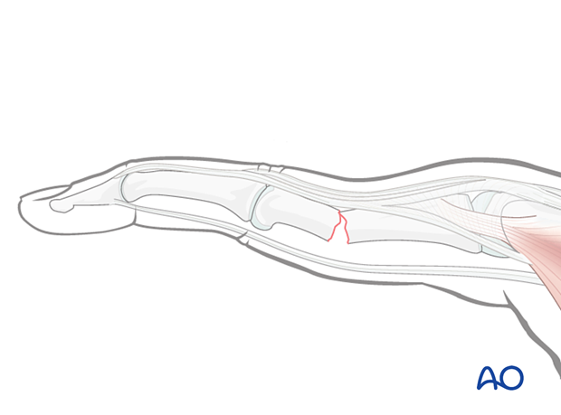
Check for shortening of a finger. Interrupted finger cascade may indicate shortening.
Check for deformity of a phalanx. In a fracture situation, the distal fragment may be pulled dorsally.
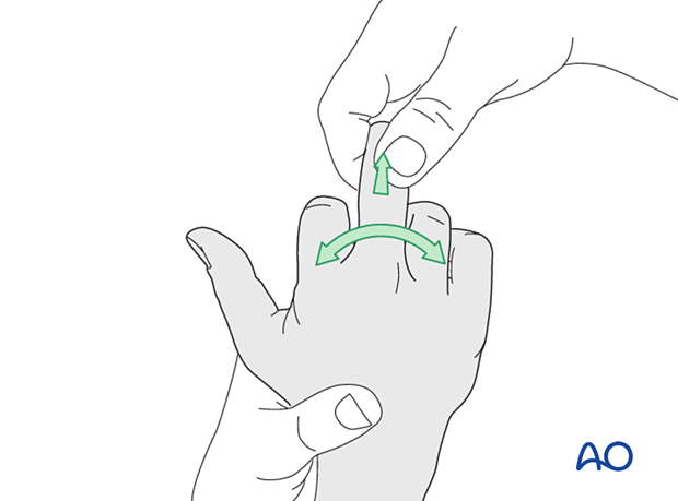
Check the metacarpophalangeal (MCP) and proximal interphalangeal (PIP) joint stability. Compare this with the contralateral finger.
Lack of lateral stability indicates injury of the collateral ligaments or their bony attachments.
3. Radiologic evaluation
X-rays
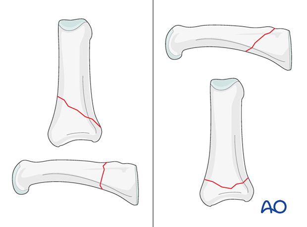
AP view of the whole hand and lateral view of the finger are needed for diagnosis. In base fractures, an oblique view may be helpful.
All views need to be inspected to get information about the fracture pattern, eg, the orientation of an oblique fracture plane.
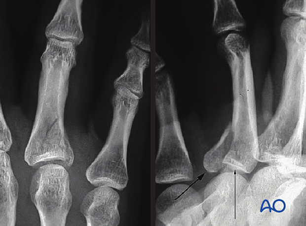
Obliquity of the fracture is possible either in the plane visible in the AP view or the plane visible in the lateral view. Always confirm the fracture configuration with views in both planes.
Ultrasound
Ultrasound evaluation may help define suspected ligament and tendon injuries.
CT imaging
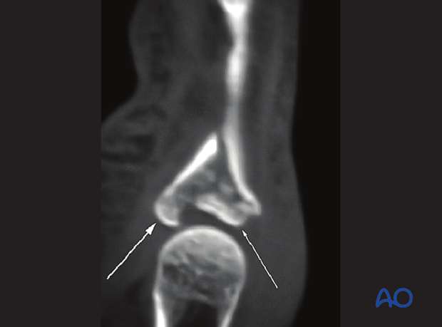
CT imaging is required for impacted intraarticular fractures to evaluate the fragmentation and plan for reconstruction.
MRI
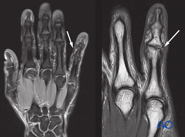
Isolated ligamentous injuries may be seen in an MRI.
