Complete articular fracture of the distal end segment
Definition
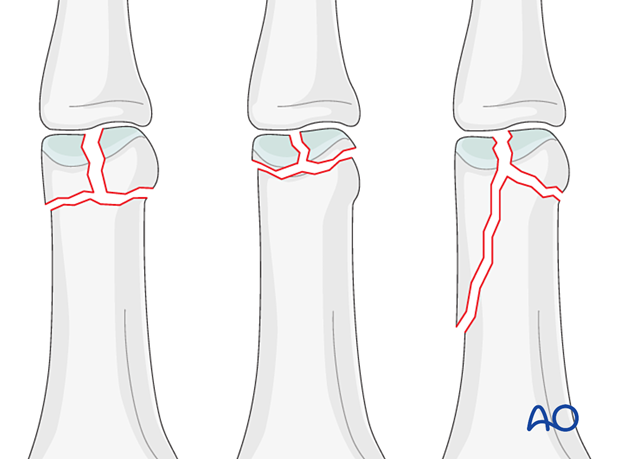
Bicondylar articular fractures of the proximal phalangeal head are classified according to AO/OTA as 78.2–5.1.3C, where 2–5 indicates which finger is injured.
These fractures may be T- or Y-shaped, with a long or a short T. Another pattern of fracture is a combination of a long oblique fracture separating one condyle, together with a short oblique, or transverse, fracture separating the other condyle (sometimes called “reversed lambda” fractures, because of their resemblance to the Greek letter λ).

Further characteristics
Typically, these fractures result from sports injuries due to axial load combined with lateral angulation of the finger.
Condylar fractures tend to be very unstable because the ligaments usually pull at the fragments and displace the fracture.
Imaging
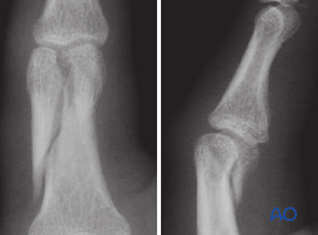
AP and lateral view x-rays of a bicondylar fracture with an extension into the diaphysis

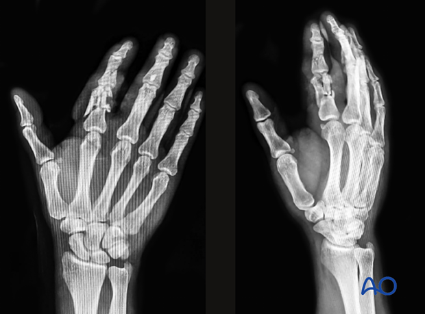
AP and oblique view x-rays of a multifragmentary articular fracture of the 2nd proximal phalangeal head and a fracture of the middle phalanx

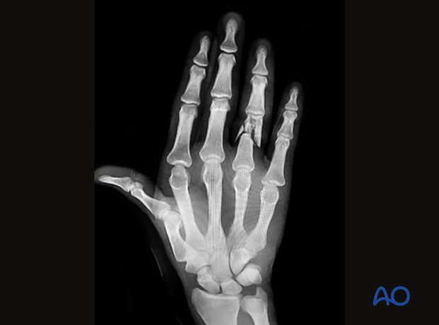
AP view x-ray of a multifragmentary articular fracture of the 4th proximal phalangeal head


