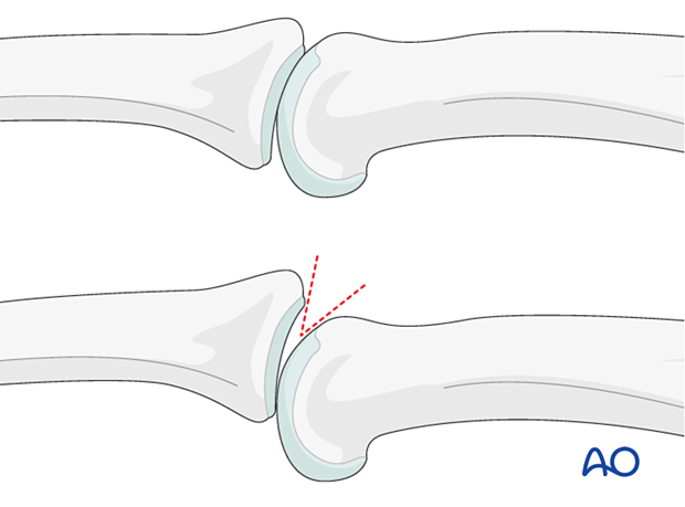
Multifragmentary articular fracture of the proximal end segment
Definition
Multifragmentary articular fractures of the middle phalangeal base are partial or complete articular fractures and classified according to AO/OTA as 78.2–5.2.1B or 1C, respectively, where 2–5 indicates which finger is injured.
These fractures may be associated with a proximal interphalangeal (PIP) joint dislocation.
The following fracture types are described:
- Central and palmar impaction fracture
- Lateral plateau compression fracture
- Complete articular fracture
- Pilon compression fracture
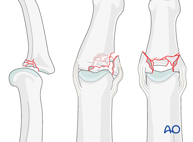
Central and palmar impaction fracture
The degree of comminution is often not apparent on the x-rays. These fractures are usually associated with impaction in the subchondral metaphysis.
With increasing impaction and comminution, the stability of the joint decreases.
- <15%: stable
- 15–30%: mildly unstable
- >30%: unstable
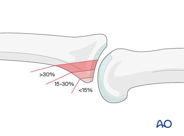
Impaction is possible both in the sagittal and the coronal planes.
Impaction can occur in several areas, either involving the volar ring, up to 50% of the palmar joint aspect, or the central section of the joint.
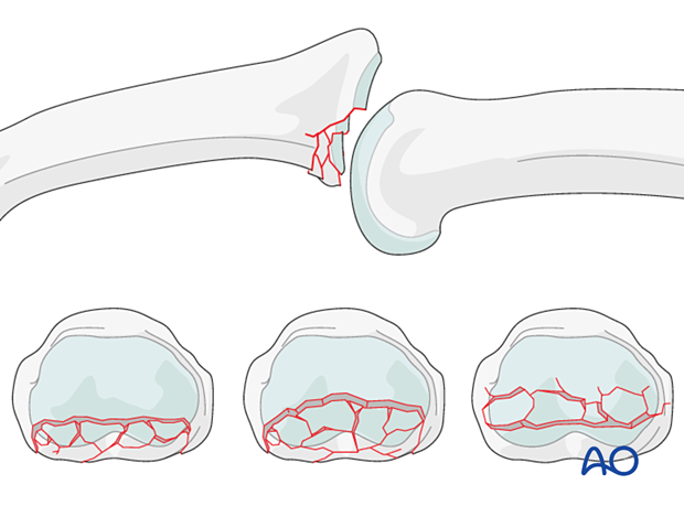
Mechanism of the injury
These digital hyperextension injuries are commonly caused by sporting accidents.
Typically, hyperextension of the proximal interphalangeal (PIP) joint causes an avulsion fracture of the volar plate.
Often, in addition to hyperextension, impact on the fingertips causes longitudinal compression forces through the middle phalanx proximally, leading to an additional impaction fracture of the proximal phalangeal head.

Deforming forces
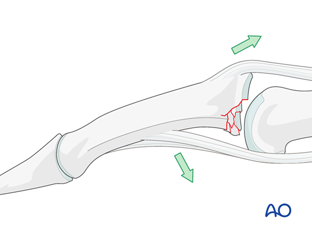
The flexor digitorum superficialis (FDS) exerts a palmar pull on the middle phalanx around a pivotal point determined by the junction of intact cartilage and the fracture; this leads to subluxation and dorsal tilting, depending on the degree of the impaction.
In the presence of palmar instability of the PIP joint, muscle forces (flexor digitorum superficialis and the central extensor slip) lead to palmar tilting and dorsal subluxation, depending on the degree of the impaction.
Imaging
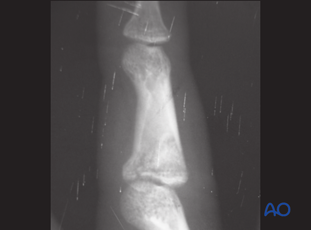
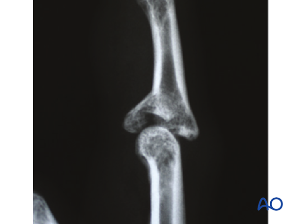
This lateral x-ray shows a palmar impaction fracture with minimal displacement.
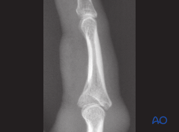
This lateral x-ray shows a palmar impaction fracture with depression of the articular surface.
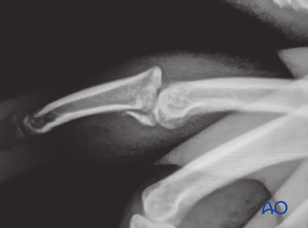
This lateral x-ray shows a palmar multifragmentary impaction fracture with dorsal dislocation.
This lateral x-ray shows a central impaction fracture.
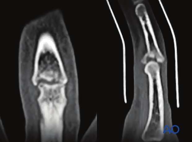
CT scan
Coronal and sagittal CT views of the case shown above
Lateral plateau compression fracture
Mechanism of the injury
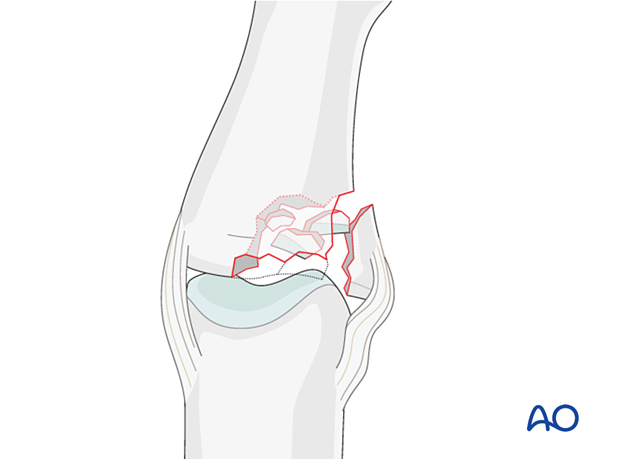
A combination of lateral angulation and axial loading of the finger results in eccentric longitudinal compression forces on one condyle of the middle phalanx, leading to impaction fracture.
Dorsal or dorsal rotatory instability may occur.
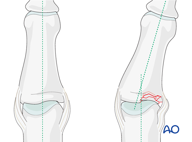
Malalignment in the coronal plane may be a sign of impaction.
Imaging
X-rays in two views are needed for assessing the fracture. For detailed assessment of the articular surface fragmentation, CT scan is helpful.
Complete articular and pilon compression fractures
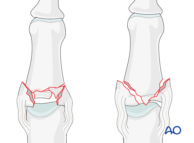
When vertical compression forces, created by axial load, are applied to the finger, comminuted, intraarticular, compression fractures may result. These fractures are very unstable.
Typically, they occur in two configurations:
- As centrally impacted, comminuted fractures
- With a T- or Y-type fracture geometry
Impaction injuries to the middle phalanx can result in a fracture dislocation of the PIP joint with variable amounts of damage to the articular surface. These injuries are generally unstable and result in joint stiffness and late development of osteoarthritis.
Imaging
X-rays in two views are needed for assessing the fracture. For detailed assessment of the articular surface fragmentation, CT scan is helpful.
Recognizing subluxation
In the lateral view, the dorsal cortical profiles of the proximal and middle phalanges should be collinear. Any axial malalignment is a clear indication of subluxation.
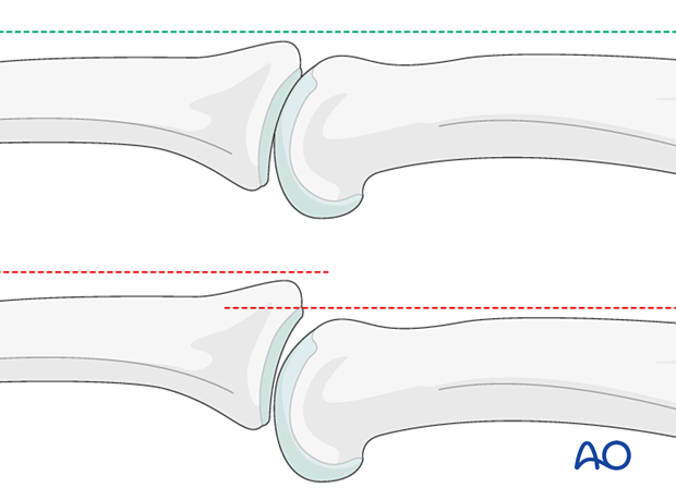
Another indication of subluxation is the presence of a so-called V-sign in the lateral x-ray.
