Extensor block pinning with joint transfixation
1. General considerations
Introduction
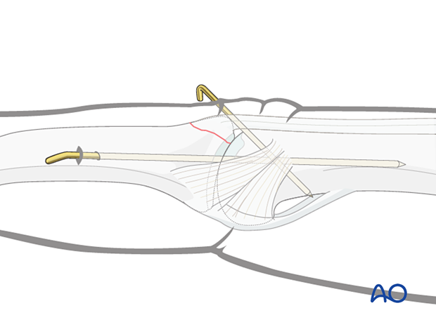
Extensor block pinning stabilizes the dorsal fragment if it remains displaced after relocation of the joint. Additionally, the PIP joint is transfixed with an axial K-wire.
Extensor block pinning can be used in case of smaller displaced dorsal fragments.
Recovery process
The recovery process after such injuries is slow. Advise the patient to expect 6–8 months for full recovery.
Potential risks of percutaneous pinning
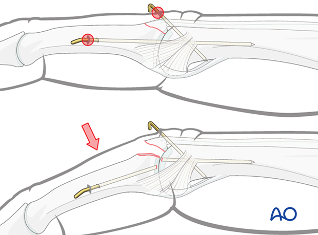
The commonest risk of percutaneous K-wire immobilization is infection.
Breakage of the K-wire at the level of the PIP joint can be avoided by using a wire of 1.2 mm diameter, splint immobilization, and counseling the patient to avoid stresses at the PIP joint.
2. Patient preparation
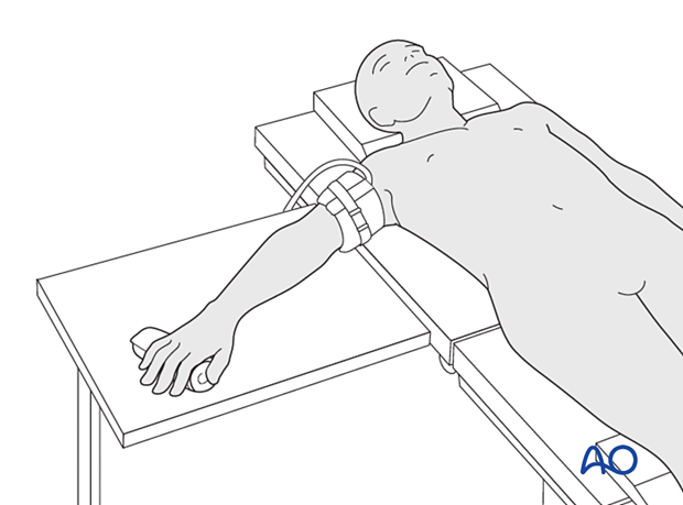
Place the patient supine with the arm on a radiolucent hand table.
3. Insertion of extension block
Determine the precise location of the PIP joint with an image intensifier.
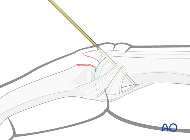
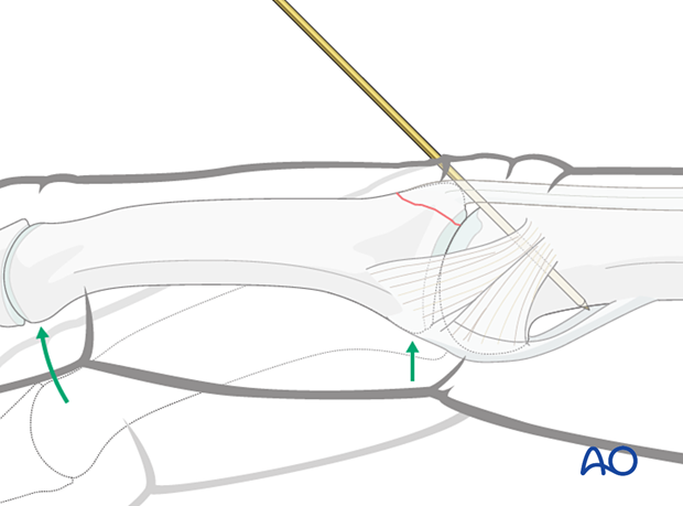
Use a drill guide to insert a 1.2 mm K-wire at an angle of approximately 45° through the central band into the head of the proximal phalanx, ensuring that the avulsion fragment is reduced. Engage the far cortex.
This K-wire serves as the extension block.
Reduction
Carefully reduce the fracture by extending the PIP joint and adjusting the position of the distal fragment.
Confirm PIP joint reduction and position of the avulsion fragment with an image intensifier in the lateral view.
4. Joint transfixation with K-wire
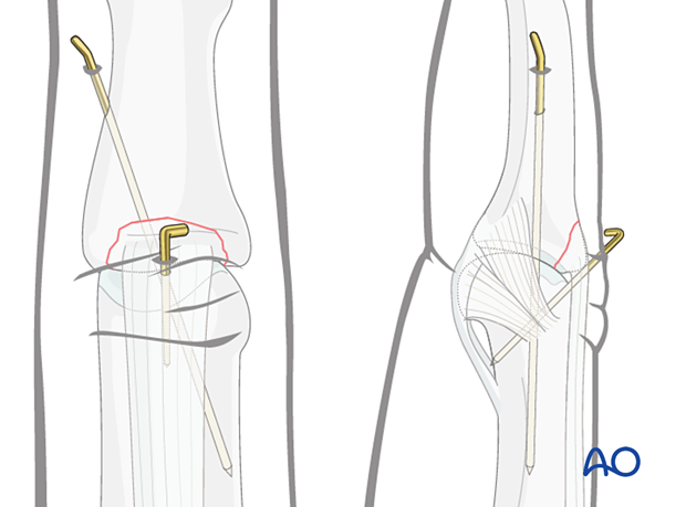
Transfixation of the joint for 6 weeks is required to protect the central slip reattachment. There is an increased risk of joint stiffness.
Pass a 1.2 mm K-wire obliquely across the PIP joint with the joint in extension.
Leave the end of the K-wire outside of the skin to facilitate later removal.
The fixation must be protected with a splint to reduce the risk of wire breakage.
5. Final assessment
Check joint congruity using image intensification. Reduction must be anatomical.
6. Aftercare
Postoperative phases
The aftercare can be divided into four phases of healing:
- Inflammatory phase (week 1–3)
- Early repair phase (week 4–6)
- Late repair and early tissue remodeling phase (week 7–12)
- Remodeling and reintegration phase (week 13 onwards)
Full details on each phase can be found here.
Postoperative treatment
If there is swelling, the hand is supported with a dorsal splint for a week. This should allow for movement of the unaffected fingers and help with pain and edema control. The arm should be actively elevated to help reduce the swelling.
The hand should be immobilized in an intrinsic plus (Edinburgh) position:
- Neutral wrist position or up to 15° extension
- MCP joint in 90° flexion
- PIP joint in extension
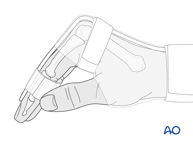
The MCP joint is splinted in flexion to maintain its collateral ligaments at maximal length to avoid contractures.
The PIP joint is splinted in extension to maintain the length of the volar plate.
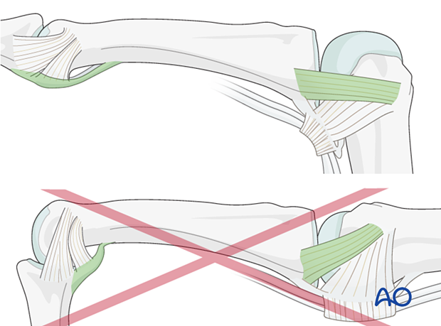
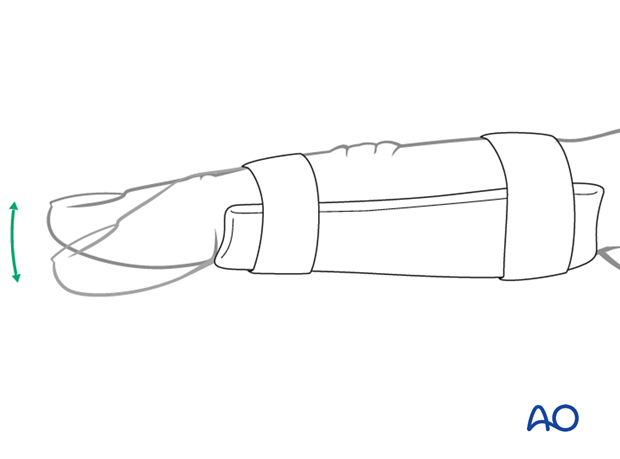
After swelling has subsided, the PIP joint is protected in extension with a palmar splint, leaving the DIP joint free.
Cleaning
Removal of the splint and skincare must be performed by the patient at weekly intervals.
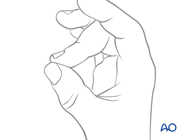
Instruct the patient to keep the finger in extension by pinching it with the thumb when the splint is taken off for cleaning or by pressing the tip of the finger onto a tabletop.
Any flexing of the finger may disrupt the healing process.
Mobilization
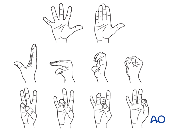
To prevent joint stiffness, the patient should be instructed to begin active motion (flexion and extension) of all nonimmobilized joints immediately after surgery.
After K-wire and splint removal, unrestricted active flexion and extension is permitted.
Follow-up
X-ray checks of joint position must be performed immediately after the splint has been applied.
Follow-up x-rays with the splint should be taken after 1 week and, if necessary, after 2 weeks. A final x-ray can be taken at the expected fracture consolidation.
In the middle phalanx, the fracture line can be visible in the x-ray for up to 6 months. Clinical evaluation (level of pain) is the most important indicator of fracture healing and consolidation.
K-wire removal
The K-wire is removed after 4–6 weeks.


