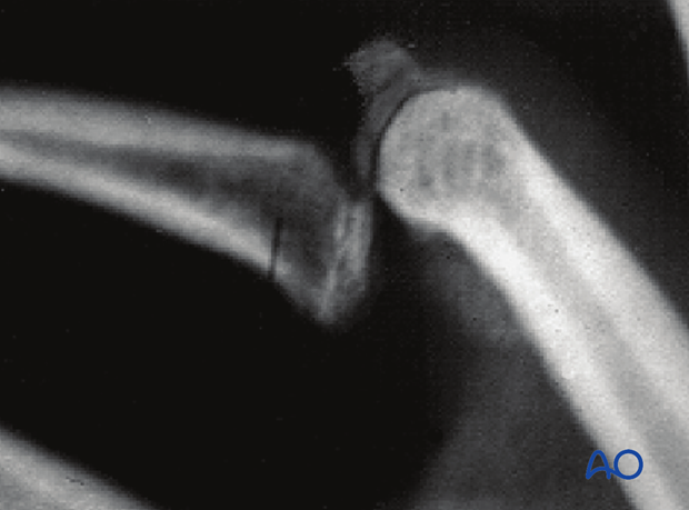
Dorsal avulsion of the proximal end segment
Definition
Avulsions involving the articular surface of the base of the middle phalanx are partial articular fractures and classified according to AO/OTA as 78.2–5.2.1B, where 2–5 indicates which finger is injured. The fractures may be simple or fragmentary.
An avulsion fracture may be associated with a proximal interphalangeal (PIP) joint dislocation.
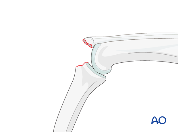
Lesion classification
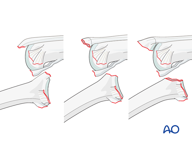
Several lesions may be produced by a hyperflexion mechanism.
All of these are characterized by detachment of the central slip of the extensor apparatus. They are considered as acute boutonnière injuries.
Lesions without dislocation
- Hyperflexion with detachment of the central slip (no fracture)
- Simple avulsion fracture of the central slip with a small nonarticular fragment
- Simple avulsion fracture of the central slip with a larger partial articular fragment
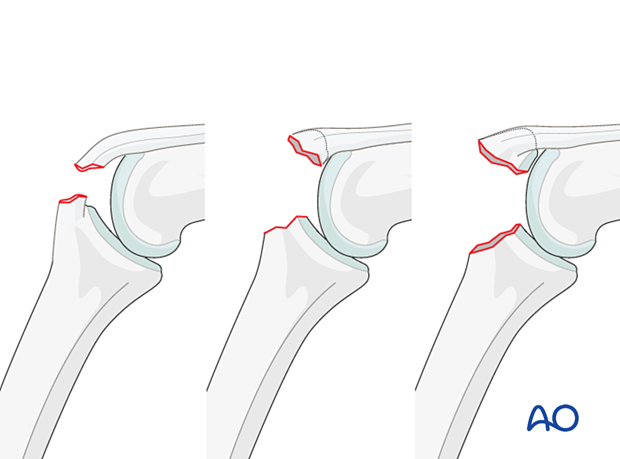
Sometimes, lesions such as these are considered minor injuries by traumatologists, and often they are confused with simple ligament sprains.
However, the boutonnière deformity and associated lesions should be addressed acutely because they are difficult to treat at a later stage.
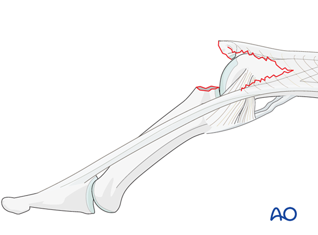
Lesions with palmar dislocation
If palmar dislocation of the proximal interphalangeal (PIP) joint is present, a rupture of the collateral ligament and detachment of the central slip are involved. In injuries of this kind, a so-called swan-neck deformity is present, and the finger can not be flexed.
Lesions with dislocations:
- Detachment of the central slip without a fracture
- Avulsion fracture of the central slip with a small nonarticular fragment
- Avulsion fracture of the central slip with a larger partial articular fragment
Mechanism of the injury
These injuries are commonly caused during sports, eg, cricket, volleyball, basketball, etc. The middle and ring fingers are commonly involved.
Typically, hyperflexion of the finger causes an avulsion lesion of the central slip.
Often, in addition to hyperflexion, impact on the fingertips causes longitudinal compression forces through the middle phalanx proximally, leading to an additional impaction fracture of the base of the middle phalanx or proximal phalangeal head.
Deforming forces
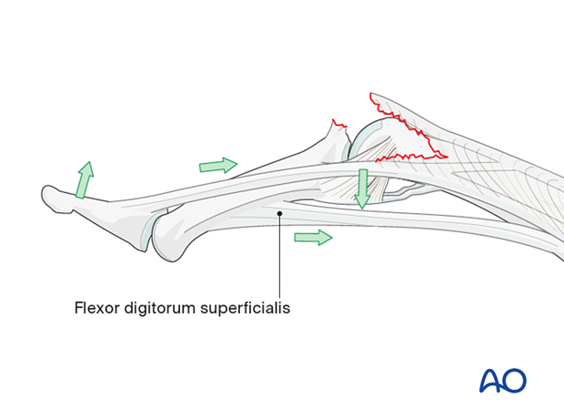
Boutonnière deformity
When the central slip is detached, the lateral bands are palmarly displaced and pull the DIP joint into hyperextension.
This creates a lesion like a buttonhole (“boutonnière”) in the extensor mechanism through which the head of the proximal phalanx perforates dorsally.
The flexor digitorum superficialis (FDS) pulls proximally on the middle phalanx, forcing the PIP joint into flexion.
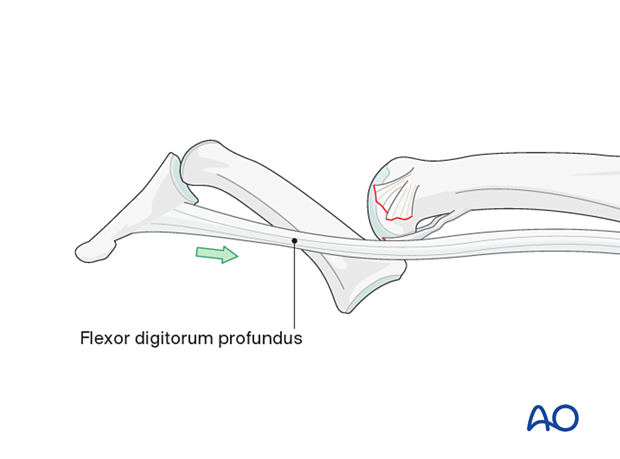
Swan-neck type deformity
If the middle phalanx is palmarly dislocated at the PIP joint by the energy of the trauma, the flexor digitorum profundus (FDP) pulls the DIP joint into flexion. This creates the characteristic deformity of the finger described as having the profile of a swan’s neck.
Recognizing subluxation
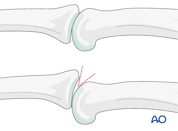
In the lateral view, the dorsal cortical profiles of the proximal and middle phalanges should be collinear. Any axial malalignment is a clear indication of subluxation.
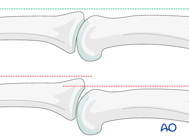
Another indication of subluxation is the presence of a so-called V-sign in the lateral x-ray.
Imaging
Lateral x-ray of a dorsal avulsion fracture with palmar dislocation
