Authors of section

Authors

Fiesky Nuñez, Renato Fricker, Matej Kastelec, Terry Axelrod

Executive Editor

Chris Colton

Proximal, collateral ligament avulsion

Classification of avulsion fractures of the volar plate

Avulsion fractures of the volar plate are very common injuries, often resulting from sporting injuries and usually involving the middle and ring fingers. Several classification systems for them have been proposed for them. The Eaton classification is useful and practical. Its premise is that successful treatment must be based on the stability of the fracture, which in turn depends on:

  • size of the fragment
  • degree of impaction
  • ruptures of one or both collateral ligaments
  • direction of the dislocation (palmar, lateral, or dorsal)

Eaton type I (hyperextension)
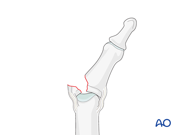
These are hyperextension injuries, with an avulsion of the volar plate and a longitudinal split in the collateral ligaments.

Eaton type II (dorsal dislocation)
Complete dorsal dislocation of the PIP joint and avulsion of the volar plate. The base of the middle phalanx rests dorsally on the condyles of the proximal phalanx, with no contact between the articular surfaces.

Eaton type III (fracture dislocation)
A fracture dislocation with an avulsed small fragment.

definition
definition

Stability of fracture dislocations (Eaton type III)

Stability of the reduction depends on the size of the avulsed fragment and the amount of ligament remaining attached to the middle phalanx.
If less than 40% of the articular segment is avulsed, the fracture is displaced dorsally, with the dorsal portion of the collateral ligament remaining attached to the middle phalanx. This helps to keep the reduction stable.
However, if more than 40% of the articular segment has avulsed, only very little or no ligament will remain attached to the base of the middle phalanx, rendering the reduction unstable.

joint reconstruction with bone graft

Mechanism of the injury

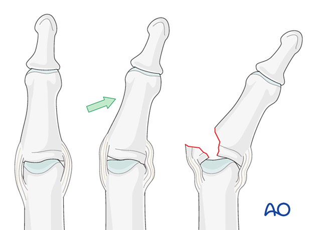
These injuries are commonly caused by hyperextension of the finger causing an avulsion fracture of the volar plate.
Often, in addition to hyperextension, axial pressure applied to the fingertip, causes longitudinal compression forces on the middle phalanx towards the proximal phalanx; this can lead to an additional impaction fracture.

lag screw fixation of volar plate

Deforming forces

The flexor digitorum superficialis (FDS) exerts a palmar pull on the middle phalanx around a pivotal point determined by the junction of intact cartilage and the fracture; this leads to subluxation and dorsal tilting, depending on the degree of the impaction.

lag screw fixation of volar plate

Recognizing subluxation

Diagnosis is based on:

  1. the clinical history of the trauma and mechanism of the injury
  2. the clinical examination of the patient
  3. the x-rays

AP and lateral x-rays are necessary for diagnosis. Be careful to avoid overlap of other fingers in the x-rays.
An AP view will help to recognize impaction fractures.
Often, a subluxation is not immediately apparent on the lateral view. Look for the characteristic “V” sign of diverging joint surfaces, which indicates this injury.

In the lateral view, the dorsal cortical profiles of the proximal and middle phalanges should be collinear. Any axial malalignment is a clear indication of subluxation.

lag screw fixation of volar plate

Indication for nonoperative treatment

Ask the patient to flex the finger under image intensification.
If reduction of the avulsion fracture is achieved with less than a 30 degree bend, nonoperative treatment may be a good choice.
However, if it takes more than 30 degrees of flexion to reduce the fragment, operative treatment is indicated.
Reduction will not be achieved if soft tissues are interposed between the fracture fragments. This is also an indication for surgical treatment.
Passive lateral movement of the finger under image intensification will help to assess lateral stability.

definition

Small-fragment avulsion fracture

When the avulsed fragment is very small (<30% of the articular segment), nonoperative treatment is usually a good choice.

definition

Irreducible fracture – operative treatment

When the avulsed fragment is very small (<30% of the articular segment), nonoperative treatment is usually a good choice. In some cases, the fracture can not be reduced due to interposed tissues, or blood clot.
In these cases, internal fixation is indicated.
As the use of a lag screw could easily shatter such a small fragment, internal fixation with sutures is often chosen.

definition
Go to indication