Partial articular fracture of the distal end segment
Definition
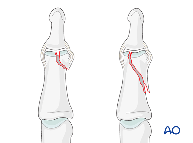
Oblique articular (unicondylar) fractures of the middle phalangeal head are classified according to AO/OTA as 78.2–5.2.3B, where 2–5 indicates which finger is injured.
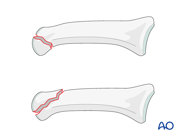
Unicondylar fractures of the middle phalanx can be oblique (short or long) or comminuted.
Short oblique fractures usually originate in the intercondylar notch.
Long oblique fractures more often pass through one of the condyles, splitting proximally towards the diaphyseal cortex on the side of the uninjured condyle.
Typically, they are the results of an axial load combined with lateral angulation of the finger.
Coronal fractures of the distal end segment are extremely rare and usually associated with a distal interphalangeal (DIP) joint dislocation.
Further characteristics
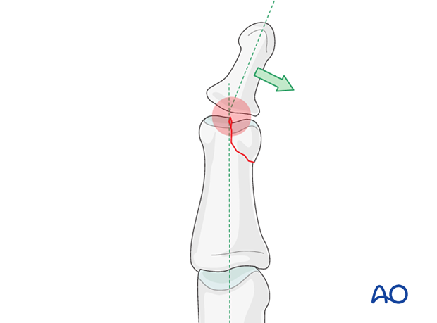
Articular fractures should be reduced anatomically. Otherwise, the articular cartilage may be damaged, leading to painful degenerative joint disease and digital deformity.
This illustration shows how minimal unicondylar depression may lead to angulation of the finger.
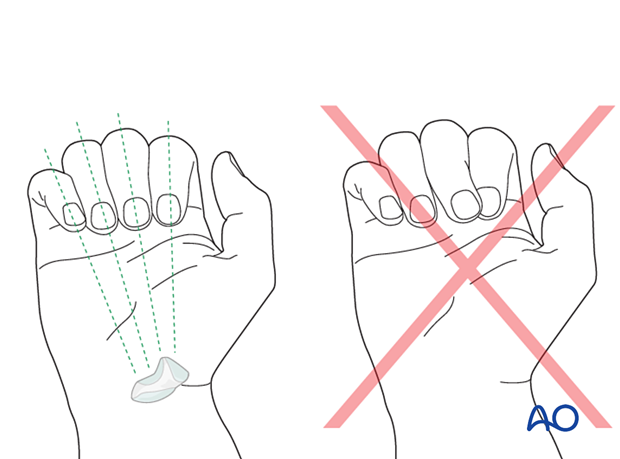
Rotational deformity
In fractures distal to the proximal interphalangeal (PIP) joint, overlap of neighboring fingers may occur when there is substantial rotational displacement.
Outcomes
Outcome of fractures of the middle phalanx is usually more favorable than those of the proximal phalanx. This is largely because limitations in distal interphalangeal (DIP) joint motion are not such a disability as similar stiffness of the PIP and metacarpophalangeal (MCP) joints.
Imaging
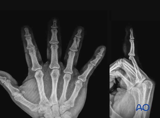
These AP and lateral x-rays show a long partial articular fracture of the 4th middle phalangeal head.
