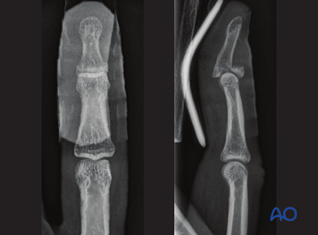
Dorsal avulsion injury of the proximal end segment (mallet finger)
Definition
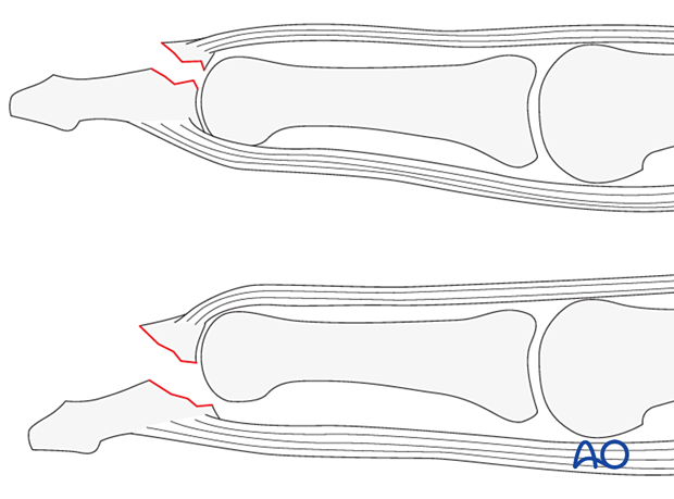
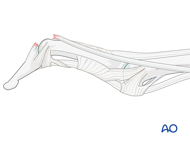
Discontinuities of the extensor insertion are often referred to as “mallet injury” or “baseball finger.” They can be purely tendinous or bony avulsion fractures.
Bony avulsions (partial articular) of the distal phalangeal base are classified by the AO/OTA as 78.2–5.3.1B, where 2–5 indicates which finger is involved.
Large bony avulsions often are associated with palmar joint dislocation.
Further characteristics
The injuries can result in subluxation or total dislocation of the joint.
A large fragment remains minimally displaced because the volar plate attachment, the collateral ligament, and the A4 pulley remain largely intact.
An avulsion injury destroys the synergistic balance of the pull exerted by the flexor and extensor tendons. The continuity of the flexor tendon is lost. This results in an inability to flex the DIP joint.
Mechanism of injury
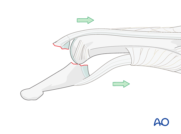
Flexion injuryThe commonest cause of these injuries is forcible flexion of the actively extended DIP joint, as when stubbing a straight finger against resistance.
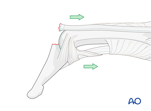
Occasionally, the injury results from an axial overload of the terminal segment of the finger, causing joint impaction and a dorsal marginal fracture, which is retracted by the pull of the extensor tendon.
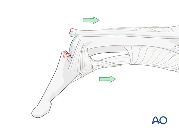
An obliquely orientated axial compression force sometimes results in a dorsal marginal fracture, involving approximately half the articular surface, and can disrupt the collateral ligaments.
Presentation of injury
Partial tendon disruptionIn incomplete tendon injuries, the resulting extension lag is no greater than 30°. The patient retains a partial ability actively to extend the DIP joint.
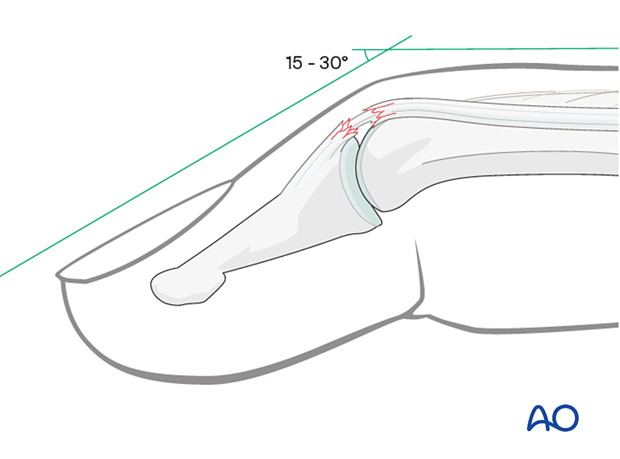
In complete disruption of the central part of the extensor mechanism, the patient is unable to actively extend the DIP joint.
The flexor digitorum profundus exerts a flexion deforming force on the distal phalanx, partly counterbalanced by the intact oblique retinacular ligaments and the collateral ligaments.
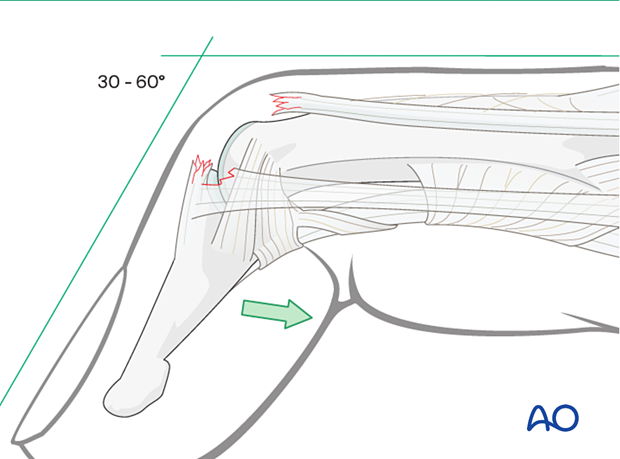
A similar clinical picture is presented by bony avulsion of the extensor mechanism at its insertion. The dorsal avulsion fracture is of variable size.
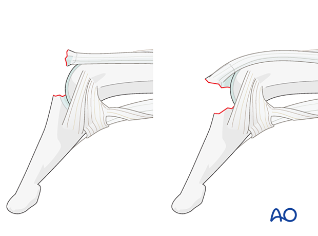
In some patients, the elasticity of the ligaments and a lax PIP joint can result in swan-neck deformity because, after disruption of the extensor mechanism at the DIP joint, all extensor forces are concentrated on the PIP joint via the middle extensor slip.
Imaging
In this case, the pull of the flexor digitorum profundus results in palmar subluxation of the distal phalanx. The palmar plate is commonly intact, and the collateral ligaments are partially ruptured.
