Compartment syndrome in the hand
1. Introduction
Compartment syndrome is a true surgical emergency.
It is caused by increasing tissue pressure which prevents capillary blood flow leading to ischemia in muscle and nerve tissue.
If not treated, tissue necrosis with permanent loss of function may occur.
Compartment syndrome may occur as a result of:
- High-energy limb injuries
- Crushing injuries
- Reperfusion injury
- Burns
- Infection
Treatment of compartment syndrome in the hand requires surgical release of the closed muscular-fascial compartments.
2. Definition
Compartment syndrome is characterized by a rise in pressure within a closed fascial compartment, sufficient to prevent effective capillary perfusion in muscle and nerve tissue.
Normal tissue pressure is 0–10 mm Hg. The capillary filling pressure is essentially diastolic arterial pressure. When tissue pressure approaches the diastolic pressure, capillary blood flow ceases.
3. Diagnosis
Symptoms
The diagnosis of compartment syndrome is clinical, and requires:
- A high index of suspicion
- Appreciation of progressive symptoms which include worsening pain with increasing analgesia requirement, paresthesia, progressive loss of sensation, progressive loss of power
The diagnosis is difficult in patients with:
- Head injury
- Loss of consciousness for other reasons
- High spinal injury
- Regional nerve blockade
- Other distracting injuries
Signs
The signs of an evolving compartment syndrome include:
- Tenderness and swelling of the affected compartment
- Increase in pain with passive muscle stretching
- Compartmental muscle weakness
- Later, sensory disturbance in the distribution of nerves traversing the compartment
- Later, weakness of muscles innervated by nerves traversing the compartment
4. Principles
General treatment principles
Effective management of an impending or established compartment syndrome requires:
- Recognition of the risk of, or actual compartment syndrome (symptoms and signs)
- Understanding the pathophysiology
- Recognition of the importance of immediate surgical treatment
- Intracompartmental pressure measurement in patients that cannot provide a reliable physical examination
- Resources to manage the aftercare and rehabilitation requirements
Pathophysiology
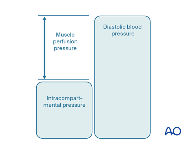
The most reliable measure of critical intracompartmental perfusion is the muscle perfusion pressure (MPP).
MPP is equal to the difference between diastolic blood pressure (dBP) and measured intramuscular pressure.
This difference in pressure reflects tissue perfusion more reliably than absolute intramuscular pressure.
When the muscle perfusion pressure is reduced to a level at which no capillary perfusion occurs, hypoxia leading to ischemia, and subsequent necrosis will occur.
The critical muscle perfusion pressure depends on the specific anatomical compartment affected.
Intracompartmental pressure measurement
When the clinical symptoms and signs of compartment syndrome are present, there is no benefit in measuring intracompartmental pressures, and an immediate fasciotomy should be performed.
When it is difficult to confirm the diagnosis, intracompartmental pressure measurement is helpful:
- To confirm the diagnosis
- To monitor a compartment at risk of increasing pressures
- To avoid unnecessary fasciotomy
- To measure intracompartmental pressures after decompression if symptoms persist
There are several techniques for the measurement of intracompartmental tissue pressure:
- Commercially available intracompartmental pressure device
- Large-bore needle and manometer
- Electronic strain gauge
If the necessary equipment is not available for direct intracompartmental pressure measurement, then the diagnosis must be assumed if there is reasonable clinical suspicion, and immediate fasciotomies must be performed.
Timing
Reversible ischemiaIn established muscle compartment syndrome, nerve and muscle tissue will become ischemic within less than two hours.
It is therefore of paramount importance that the intracompartmental pressure be released as an emergency intervention.
Arterial injury proximal to the compartment can also cause intracompartmental tissue ischemia without the associated early rise in intracompartmental pressure. After restoration of arterial flow, the subsequent capillary leakage may cause intracompartmental pressure to rise, leading to further ischemia. This is known as reperfusion injury.
It is generally accepted that after 6–8 hours of inadequate muscle perfusion pressure (MPP), extensive muscle necrosis is inevitable. Release of the muscle compartments involved will not prevent severe muscle contracture.
Fasciotomy of compartments within which muscle necrosis has already happened has a high risk of infection.
Amputation may be required.
Ischemic muscles eventually become atrophic and contracted. The movement of joints proximal and distal to the affected muscular compartment will be limited. The limb distal to the affected compartment may be insensate. The function of the limb is inevitably insufficient. This is known as “Volkmann’s ischemic contracture”.
5. Compartmental anatomy
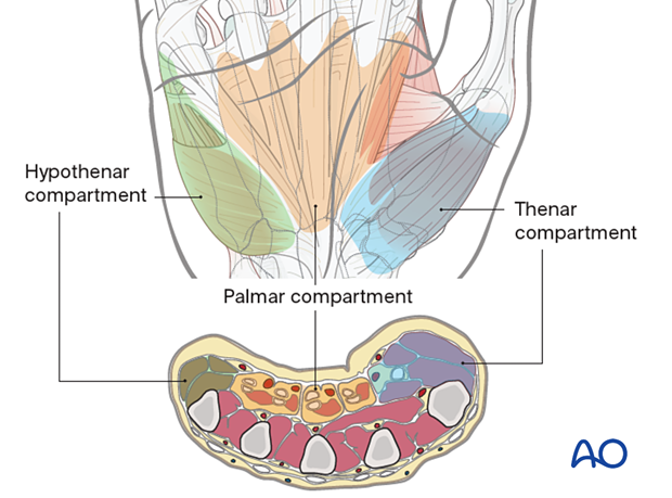
There are three main compartments in the hand separated by strong fascial septa:
- Hypothenar compartment, containing the short muscles of the little finger
- Palmar compartment, containing the median nerve, the ulnar nerve, and the long tendons to the fingers
- Thenar compartment, containing the short muscles of the thumb
6. Approaches to fasciotomy
Each of the relevant compartments must be decompressed fully by fasciotomy, using an extensile surgical approach.
If two or more compartments need to be decompressed, a combined approach may be used.
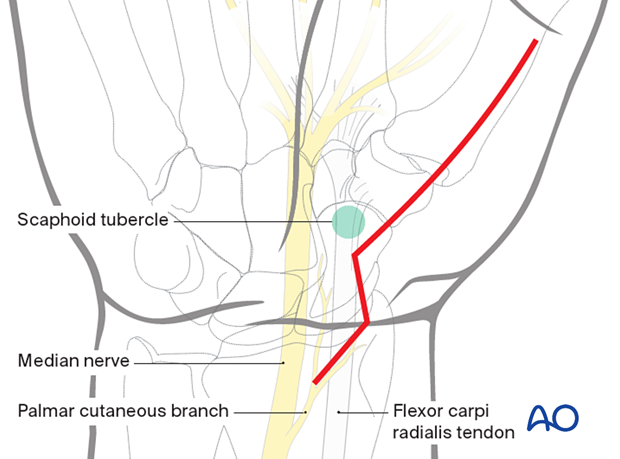
Extensile approach to the thenar compartment
For decompression of the thenar compartment an extended palmar approach to the scaphoid is used.
Care should be taken to avoid surgical injury of the palmar cutaneous branch of the median nerve.
Extensile approach to the hypothenar compartment
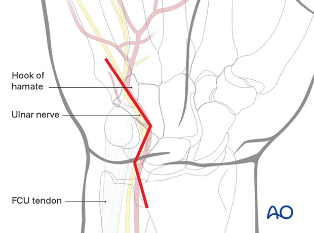
For decompression of the hypothenar compartment, an extended ulnar approach to the hamate hook may be used.
Care should be taken to avoid surgical injury of the ulnar nerve in Guyon's canal.
Extensile approach to the palmar compartment
For decompression of the palmar compartment, an extended carpal tunnel approach may be used.
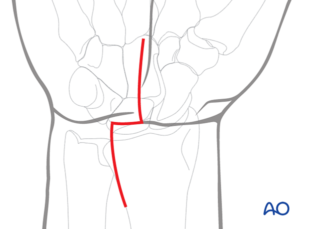
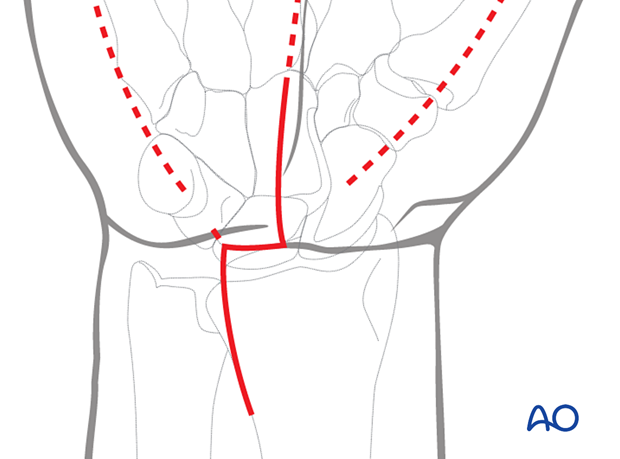
If more than one compartment of the hand is involved, particularly in the case of multiple compartment syndrome caused by extensive infection, separate incisions over each of the compartments are indicated.
Additionally, incisions over the index and ring metacarpals on the dorsal aspect of the hand facilitate decompression of the intrinsic muscles of the hand, including the lumbrical muscles.
7. Fasciotomies
Temporary soft-tissue management
After a fasciotomy or fasciotomies have been performed, skin edges retract and can become difficult to close. Temporary coverage of the wounds can be obtained with either a wound vacuum-assisted closure (VAC) device or with saline-soaked gauze bandages. These dressings or the wound VAC can be kept on until the patient returns for an attempt at secondary closure.
Delayed soft-tissue management: primary suture
If the swelling of the hand adequately decreases upon subsequent return to the operating room, primary closure of the fasciotomy wounds can occur. It is important to not perform primary closure if there is any concern about persistent swelling; secondary coverage options exist. Repeat application of an incisional wound VAC can enhance wound healing.
Delayed soft-tissue management: secondary cover
If persistent swelling exists but wound closure is necessary, particularly for associated fractures that have been fixed, secondary wound coverage options are necessary. These include split thickness skin grafting, muscle flaps, or musculocutaneous flaps. In many instances continued use of wound VACs may be sufficient for wound healing.
8. Aftercare
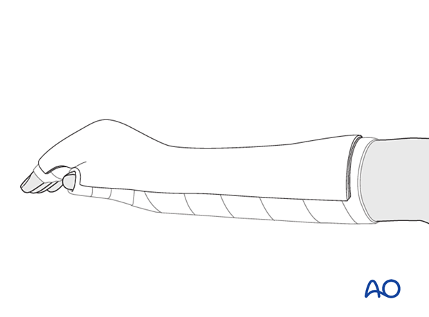
Splintage
Further information on splint application in the hand can be found here.
Rehabilitation
Active range of motion of the hand is performed as soon as possible as comfort permits. Passive mobilization is performed by physiotherapists if the patient is unable to participate.


