Compartment syndrome in the foot
1. Introduction
Compartment syndrome is a true surgical emergency.
It is caused by increasing tissue pressure which prevents capillary blood flow, leading to ischemia in muscle and nerve tissue.
If not treated, tissue necrosis with permanent loss of function may occur.
Compartment syndrome may occur as a result of:
- high-energy limb injuries
- crushing injuries
- reperfusion injury
- burns
Compartment syndrome occurs in:
- fascial compartments below the elbow
- fascial compartments below the knee
- rarely, above the elbow and knee
Treatment of compartment syndrome requires surgical release of the closed osteo-fascial compartments.
2. Definition
Compartment syndrome is characterized by a rise in pressure within a closed fascial compartment, sufficient to prevent effective capillary perfusion in muscle and nerve tissue.
Normal tissue pressure is 0–10 mm Hg. The capillary filling pressure is essentially diastolic arterial pressure. When tissue pressure approaches the diastolic pressure, capillary blood flow ceases.
3. Diagnosis
Symptoms
Diagnosis requires a high index of suspicion and appreciation of progressively severe symptoms which include the following:
- unexpected pain with increasing analgesia requirement
- paresthesia
- progressive loss of sensation
- progressive loss of power
The diagnosis is difficult in patients with:
- head injury
- loss of consciousness for other reasons
- high spinal injury
- regional nerve blockade
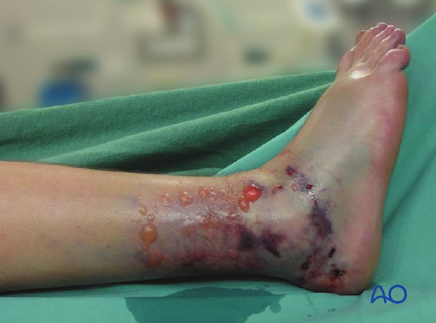
The illustration shows a distal tibial plafond fracture with swelling and bruising, resulting in fracture blisters. Compartment syndrome should be ruled out.
Signs
The signs of an evolving compartment syndrome include:
- tenderness and swelling of the affected compartment
- increase in pain with passive muscle stretching
- compartmental muscle weakness
- later, sensory disturbance in the distribution of nerves traversing the compartment
- later, weakness of muscles innervated by nerves traversing the compartment
4. Principles
General treatment principles
Effective management of an impending or established compartment syndrome requires:
- recognition of the risk of, or actual compartment syndrome (symptoms and signs)
- understanding the pathophysiology
- intracompartmental pressure measurement
- recognition of the importance of early surgical treatment
- resources to manage the aftercare and rehabilitation requirements
Pathophysiology
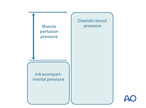
The most reliable measure of critical intracompartmental perfusion is the muscle perfusion pressure (MPP).
MPP is equal to the difference between diastolic blood pressure (dBP) and measured intramuscular pressure.
This difference in pressure reflects tissue perfusion more reliably than absolute intramuscular pressure.
When the muscle perfusion pressure is reduced to a level at which no capillary perfusion occurs, hypoxia leading to ischemia, and subsequent necrosis will occur.
The critical muscle perfusion pressure depends on the specific anatomical compartment affected.
Intracompartmental pressure measurement
When the clinical symptoms and signs of compartment syndrome are present, there is no benefit in measuring intracompartmental pressures, and an immediate fasciotomy should be performed.
When it is difficult to confirm the diagnosis, intracompartmental pressure measurement is helpful:
- to confirm the diagnosis
- to monitor a compartment at risk of increasing pressures
- to avoid unnecessary fasciotomy
- to measure intracompartmental pressures after decompression if symptoms persist
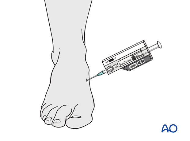
Compartment pressures should be measured at the area of maximal swelling or trauma. There are several techniques for the measurement of intracompartmental tissue pressure:
- commercially available intracompartmental pressure device
- large-bore needle and manometer
- electronic strain gauge
If the necessary equipment is not available for direct intracompartmental pressure measurement, then the diagnosis must be assumed if there is reasonable clinical suspicion, and fasciotomies must be performed.
Timing
Reversible ischemiaIn established muscle compartment syndrome, nerve and muscle tissue will become ischemic within less than two hours.
It is therefore of paramount importance that the intracompartmental pressure be released as an emergency intervention.
It is generally accepted that after 6–8 hours of inadequate muscle perfusion pressure (MPP), extensive muscle necrosis is inevitable. Release of the muscle compartments involved will not prevent severe muscle contracture.
Fasciotomy of compartments within which muscle necrosis has already happened has a high risk of infection.
Amputation may be required.
5. Compartmental anatomy
There are several foot compartments; these include:
- Lateral compartment
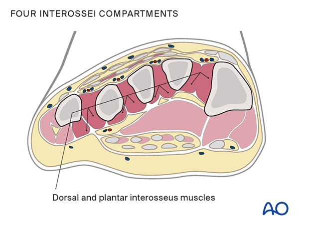
- Four interossei compartments
- Medial compartment
- Superficial compartment
- Adductor/deep compartment
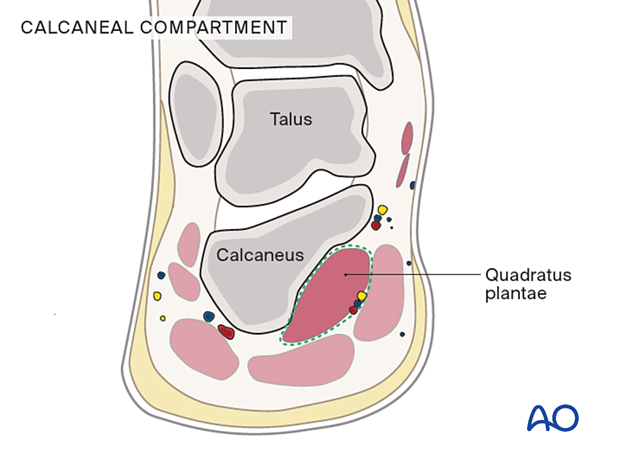
- Calcaneal compartment
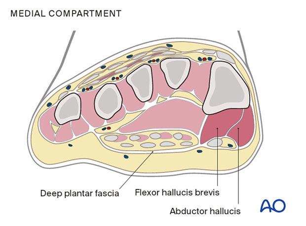
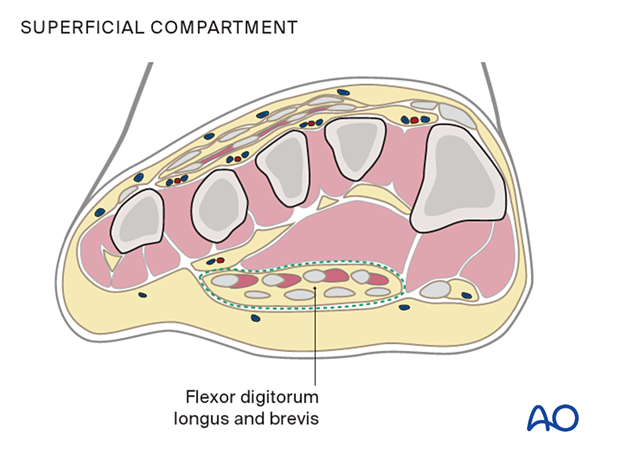
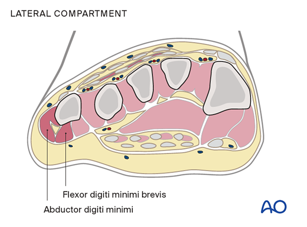
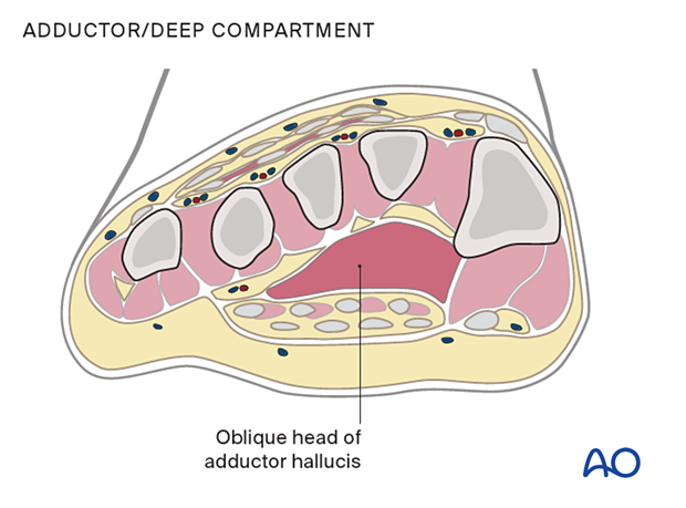
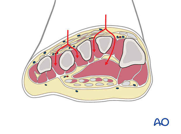
This illustration shows the compartments at the level of the forefoot.
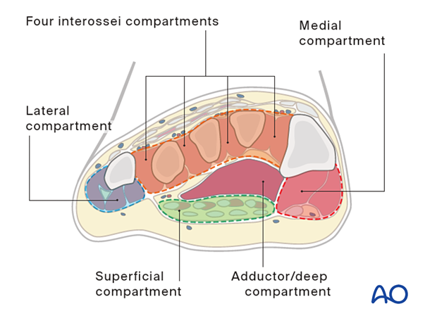
The medial compartment contains the abductor hallucis and flexor hallucis brevis muscles and is plantar-medial to the first metatarsal.
The superficial compartment contains the flexor digitorum longus and brevis muscles.
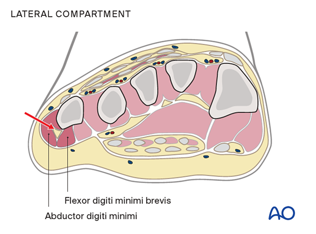
The lateral compartment contains the abductor digiti minimi and flexor digiti minimi brevis and is on the inferolateral surface of the fifth metatarsal.
The adductor or deep compartment is located in the plantar forefoot, containing the oblique head of the adductor hallucis muscle.
The four interossei compartments are dorsally located between the metatarsals, and each includes dorsal and plantar interosseus muscles.
The calcaneal compartment contains the quadratus plantae muscle.
6. General considerations
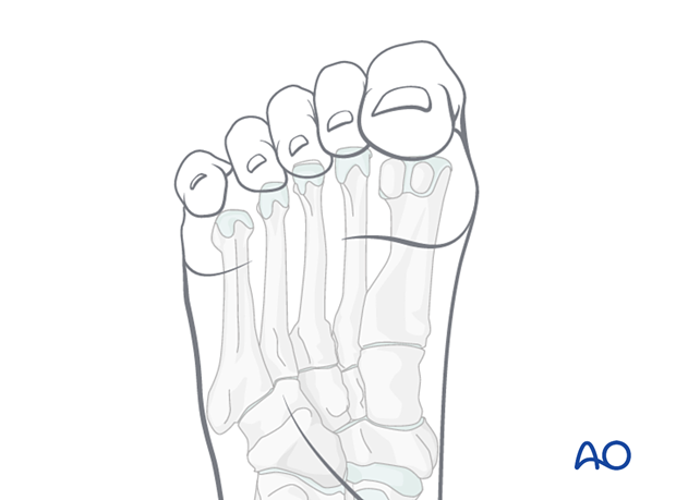
Compartment syndrome of the foot may lead to:
- Intrinsic toe contractures
- Claw toe or hammer toe
Fasciotomy may be effective in reducing the risk of intrinsic toe contractures but may lead to secondary infection and challenging soft-tissue coverage.
7. Fasciotomies
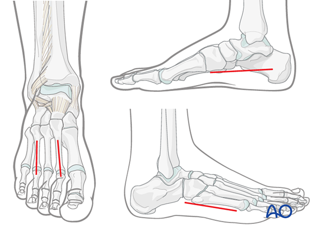
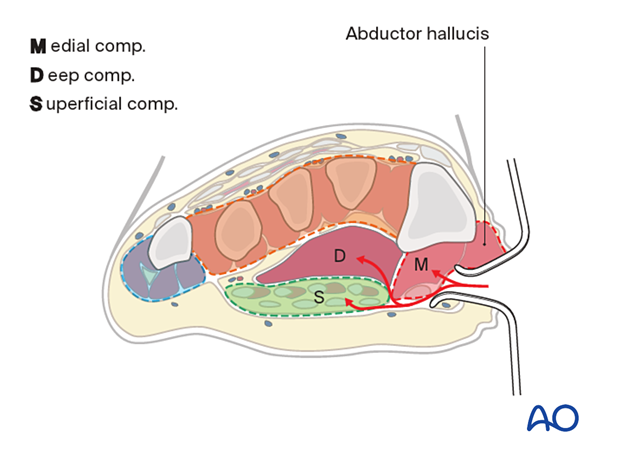
The approaches for compartment decompression generally include two dorsal incisions for access to forefoot/interossei compartments, one medial incision for decompression of the calcaneal, medial, and superficial compartments, and one lateral incision for the lateral compartments.
Double-dorsal incision
The two dorsal incisions are placed, one over the second metatarsal shaft and one over the fourth metatarsal shaft.
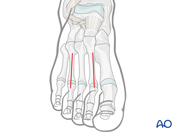
The fasciae of the interosseous muscles are opened dorsally. The muscle is stripped off the second metatarsal medially, and the fascia of the adductor compartment is opened bluntly deep within the first interspace.
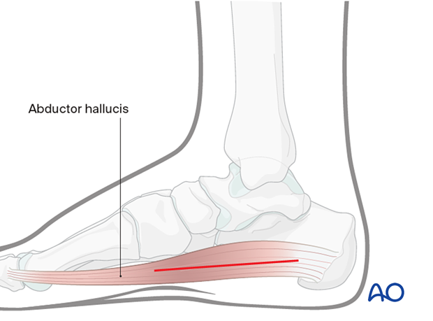
Medial incision
The medial incision is made within the foot's arch, along the muscle body of the abductor hallucis.
Dissection is continued both dorsal and plantar to the abductor hallucis muscle, which releases the medial, deep, and superficial compartments.
Lateral incision
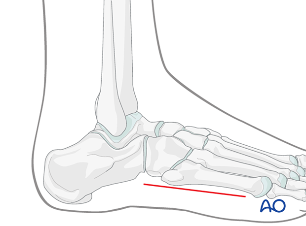
The lateral incision is made immediately plantarward of the fifth metatarsal.
The abductor digiti minimi compartment is released.
Temporary soft-tissue management
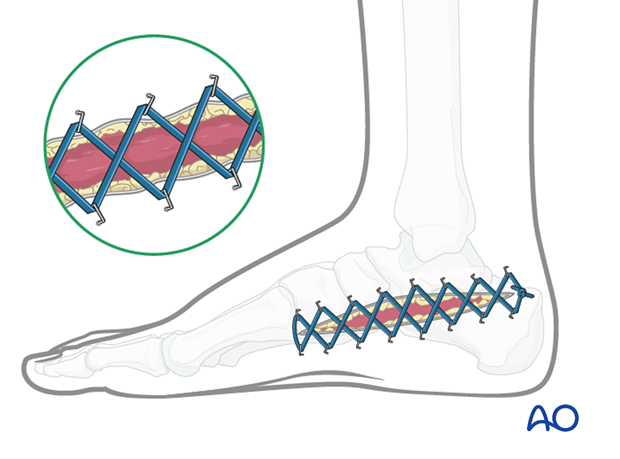
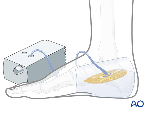
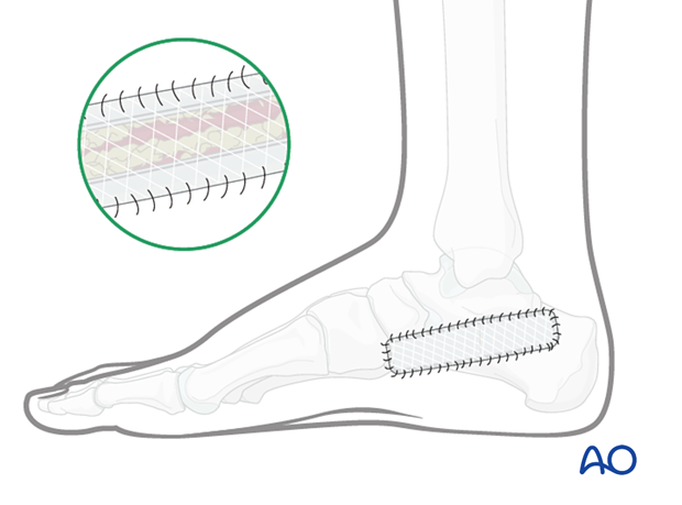
After a fasciotomy or fasciotomies have been performed, skin edges retract and can become difficult to close. Careful use of elastic retention sutures (elastic vessel loops woven through skin staples) can help counteract excessive skin contraction while still allowing the decompressed muscles to swell without any undue tension over them. Temporary coverage of the wounds can be obtained with either a wound vacuum-assisted closure (VAC) device or coverage with saline-soaked gauze bandages. These dressings or the wound VAC can be kept on until the patient returns for an attempt at secondary closure.
Delayed soft-tissue management: primary closure
If the swelling of the limb adequately decreases upon subsequent return to the operating room, primary closure of the fasciotomy wounds can occur. It is important not to perform primary closure if there is any concern about persistent swelling; secondary coverage options exist. In many instances, application of an incisional wound vac can enhance wound healing.
Delayed soft-tissue management: secondary coverage
If persistent swelling exists but wound closure is necessary, particularly for fractures that have been fixed, secondary wound coverage options are necessary. These include split thickness skin grafting, muscle flaps, or musculocutaneous flaps. In many instances wound vacs are employed to enhance wound healing.
It is imperative to cover fractures that have been fixed in a timely manner so as to minimize the risk of subsequent infection.
8. Aftercare
Splintage
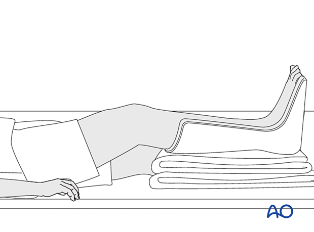
It is important to splint the foot and ankle in a neutral position to maintain a plantigrade foot, particularly if any muscle damage has occurred, as flexion contractures may develop. This can be done with a well-padded plaster back-slab, or with the extension of an external fixator to the foot. Maintain toe mobility with passive stretching.
Rehabilitation
Once the wounds have healed, assisted and active range of motion exercises are started to avoid the development of contractures. Flexion contractures of the ankle are common. Splintage in the resting position is maintained until active movement is achieved. Regular monitoring in physical therapy is maintained until optimal restoration of gait.
Late reconstruction in the presence of subsequent deformity includes the following:
- Excision of scarred muscle, tenotomies, and correction of structural deformities with tendon transfers and rebalancing
- Providing a plantigrade foot without static structural deformity
- Providing the patient with the possibility of wearing standard shoeware
9. References
General compartment syndrome references
Gourgiotis S, Villias C, Germanos S, et al Acute limb compartment syndrome: a review. J Surg Educ. 2007 64(3):178-86.
Mabee JR Compartment syndrome: a complication of acute extremity trauma. J Emerg Med.1994 12(5):651-6.
McQueen MM, Duckworth AD. The diagnosis of acute compartment syndrome: a review. Eur J Trauma Emerg Surg. 2014 Oct;40(5):521-8.
McQueen MM, Gaston P, Court-Brown CM. Acute compartment syndrome. Who is at risk? J Bone Joint Surg Br. 2000 Mar;82(2):200-3.
Powell-Bowns MF, Littlechild JE, Yapp LZ, et al. Tibial shaft fractures - to monitor or not? a multi-centre 2 year comparative study assessing the diagnosis of compartment syndrome in patients with tibial diaphyseal fractures. Injury. 2021 Oct;52(10):3111-3116.
von Keudell AG, Weaver MJ, Appleton PT, et al. Diagnosis and treatment of acute extremity compartment syndrome. Lancet. 2015 Sep 26;386(10000):1299-1310.
Compartment syndrome in the foot
Chen JS, Tejwani NC. Compartment Syndrome of the Foot. Orthop Clin North Am. 2022 Jan;53(1):83-93.
Du W, Hu X, Shen Y, et al. Surgical management of acute compartment syndrome and sequential complications. BMC Musculoskelet Disord. 2019 Mar 4;20(1):98.
Kierzynka G, Grala P. Compartment syndrome of the foot after calcaneal fractures. Ortop Traumatol Rehabil. 2008 Jul-Aug;10(4):377-83.
Lugo-Pico JG, Aiyer A, Kaplan J, et al. Foot Compartment Syndrome Controversy. 2019 Sep 3. In: Mauffrey C, Hak DJ, Martin III MP, eds. Compartment Syndrome: A Guide to Diagnosis and Management [Internet]. Cham (CH): Springer; 2019. Chapter 10.


