Minimally invasive osteosynthesis approach to the femoral shaft
1. Principles
The advantages of minimally invasive plate osteosynthesis of the femoral shaft are lessening of the extent of muscle detachment and reduced compromise of bone vascularity.
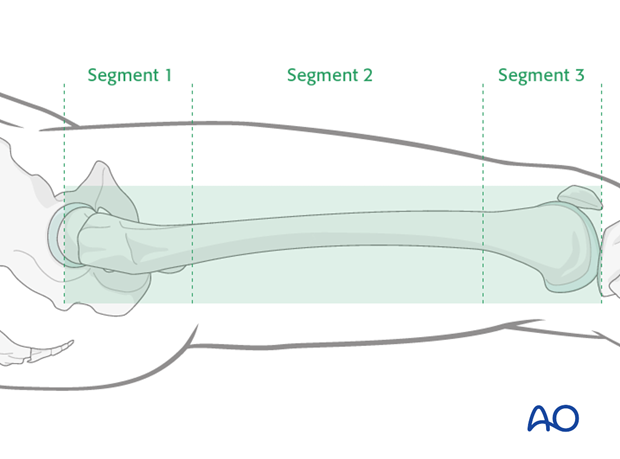
Approaches for minimally invasive plate osteosynthesis are usually performed laterally. They vary according to the fracture location: proximal (segment 1), mid-shaft (segment 2), or distal femur (segment 3) sections.
Thorough preoperative planning is compulsory in order to define the necessary specific approach locations.
2. MIO approach to the subtrochanteric region
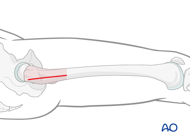
The lateral proximal incision starts at the greater trochanter and continues distally as far as needed.
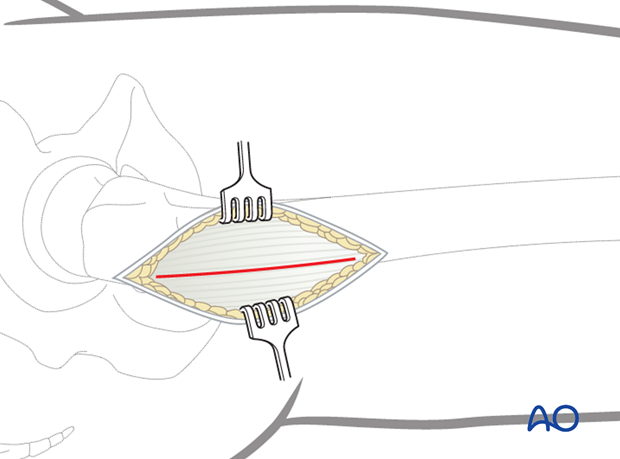
The fascia lata is incised with a scalpel and split with scissors parallel to the skin incision along the fibers.
The muscle fascia over the vastus lateralis is exposed.
If exposure of the proximal femoral shaft is necessary, the origin of the vastus lateralis must be identified.
The muscle can be retracted anteriorly with an L-shape incision down to the bone. It is then dissected off with the periosteal elevator.
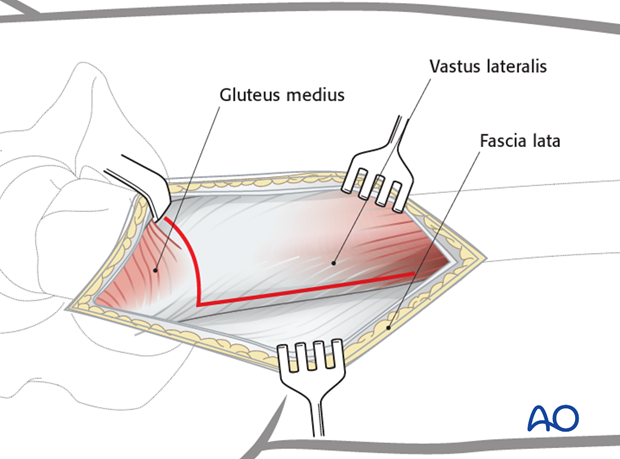
The proximal femoral shaft is exposed after performing the L-shaped extraperiosteal detachment of the vastus lateralis. The vertical part of the incision lies in the interval between gluteus medius and vastus lateralis.
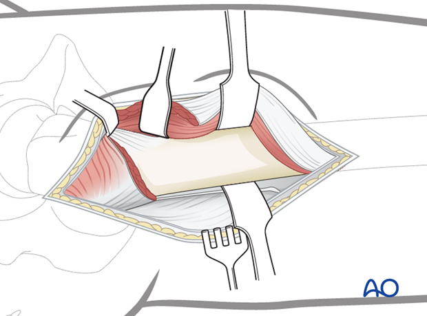
3. MIO approach to the femoral midshaft
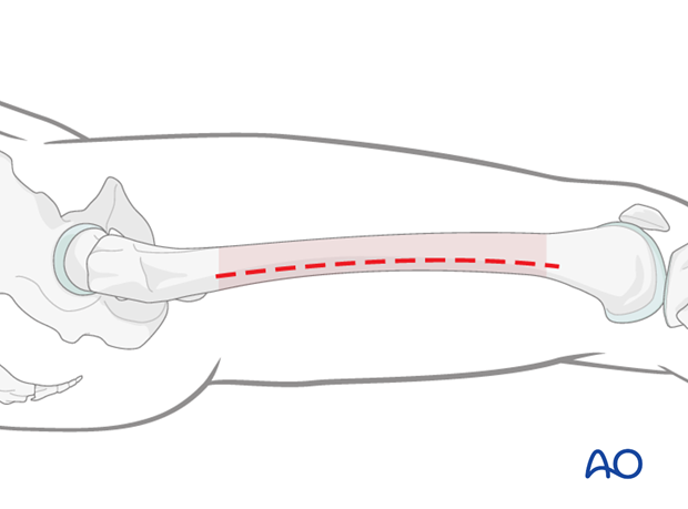
A short, or even a stab, incision is made along an imaginary line between the lateral femoral epicondyle and the greater trochanter.
The starting point and the length of the incision depend on the operational requirements for the minimally invasive procedure.
The fascia lata is incised and the muscle fascia of the vastus lateralis is exposed.
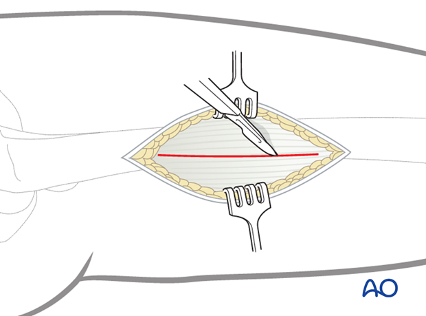
The muscle fascia of the vastus lateralis is carefully incised.
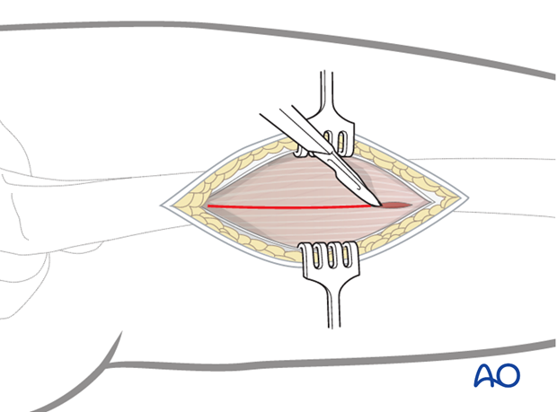
This small lateral incision provides enough access but avoids large incisions and devascularization of muscle.
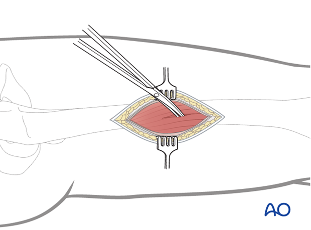
Blunt dissection is carried directly through the belly of the vastus lateralis muscle to bone.
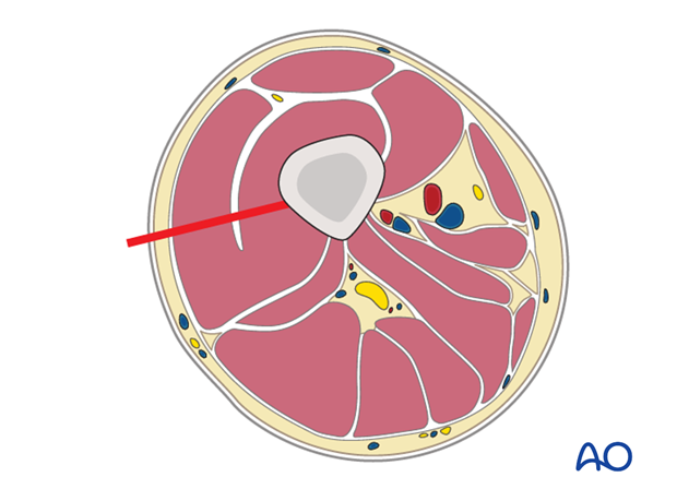
The use of two Hohmann retractors is recommended - one anterior and one posterior - for a secure exposure of the femoral shaft. These Hohmann retractors also ensure appropriate plate positioning on the femur.
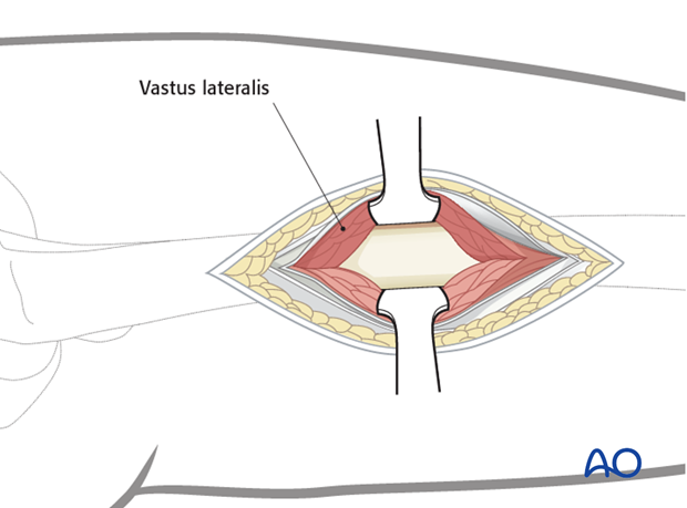
4. MIO approach to the distal femoral shaft
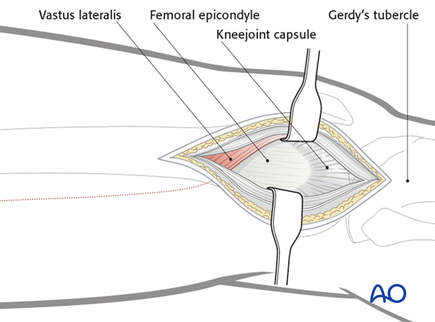
An incision is made laterally as far as the femoral epicondyle distally, directed towards Gerdy’s tubercle.
The starting location and the length of the incision can be adapted to suit operational requirements.
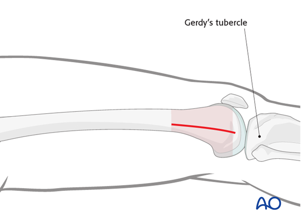
The fascia lata is incised and split along its fibers, parallel to the skin incision.
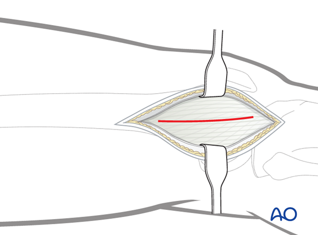
The distal femur is exposed as far as the distal margin of the vastus lateralis muscle. The plane between the femur and vastus lateralis muscle is the entry point for the plate.
