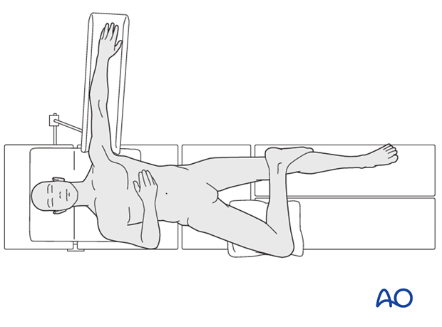
Supine position, figure-of-four
This position provides access to the posterior medial tibia. It may be used for a portion of an operation, the rest of which is done with the hip and knee extended into the standard supine position.
The patient is placed supine on a radiolucent table with the injured leg prepped circumferentially and the leg draped free.
The hip joint is abducted and externally rotated.
The knee is flexed of 90 degrees, and the foot is rested on the anterior aspect of the contralateral knee, with appropriate padding.
Blankets, or a foam pillow may be used as needed to support the knee of the injured limb. Additional external rotation of the injured limb can be obtained by placing a bump beneath the opposite hip.
A well-padded tourniquet should be applied on the thigh. Use of a tourniquet is determined by the surgeon’s preference.
The image intensifier is placed on the same side as the injured limb. It may need to be moved during fibular fracture fixation.


