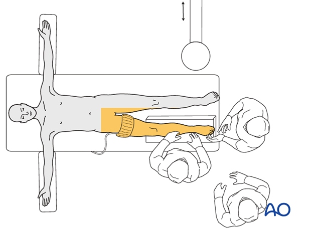
Supine position
This position allows free access to both the lateral and the medial sides by hip rotation and fluoroscopic imaging in both planes.
The patient is positioned supine on a radiolucent operating table extension with the foot brought to the end of the table. The injured leg is elevated on folded linen or a molded foam cushion. A bump consisting of a single rolled blanket is placed beneath the ipsilateral hip. This will help avoid the leg’s tendency to externally rotate.
The image intensifier is on the side opposite the injured limb.

The entire leg is prepped from toes to upper thigh. The possible need for autologous bone graft should be anticipated at this stage. If necessary, a donor site is prepared and draped.
A well-padded tourniquet should be applied to the thigh. Its inflation is determined by surgeon’s preference. Often omitted, it might be very helpful, especially during articular surface reconstruction.


