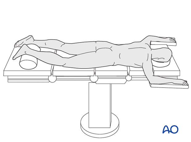
Prone position
The prone position provides access to the posterolateral and posteromedial approaches.
The patient is positioned prone on a radiolucent table with the knee flexed to 20°, padding under the thigh, and the ankle placed over a bump. It is important to ensure the foot is hanging free over the bump to aid reduction of any displaced posterior fragment and to avoid excessive pressure on the toes.
The image intensifier is positioned on the opposite side of the table, so that the C-arm can be swung underneath the table for lateral imaging. Be certain the normal leg does not interfere with fluoroscopic imaging.
A tourniquet should always be applied to the thigh but not necessarily be inflated, depending on the surgeon’s preference.
Note: Improve the preoperative preparation with the WHO Surgical Safety Checklist.


