Assessment of reduction of type A distal tibial fractures
1. Principles
During the different steps of the operation, the reduction has to be assessed repeatedly using visual and fluoroscopic control. Angulation, length of both tibia and fibula, and rotation need to be considered, as does the integrity of the ankle mortise.
Rotation is difficult to assess radiographically. By physical exam, one should confirm rotational symmetry with the opposite side.
There is a considerable variation of anatomy in the ankle region, regarding tibial shaft alignment, length of the fibula in relation to the medial malleolus, and width of the syndesmosis. Therefore, reference should be made to radiographs of the intact opposite side. Intraoperative fluoroscopy of the opposite (uninjured) leg may also be helpful.
Acceptable reduction:
Axial alignment: <5º of varus or valgus and <10º of anterior or posterior angulation. Malrotation of more than 5 or 10º may significantly interfere with certain pursuits (alpine skiing), but interferes minimally with usual activities of daily living.
Fibular length and width of syndesmosis: <2 mm difference to the opposite, uninjured side.
2. Articular surface
The tibiotalar joint lines should be parallel on the entire length in the AP as well as in the lateral view.

3. Axial alignment
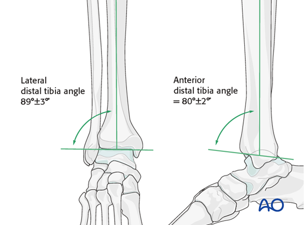
Tibial shaft alignment varies considerably. The medial distal tibia joint angle varies between 88 to 94° in the AP, and the anterior distal tibial joint angle between 78 to 82° in the lateral view.
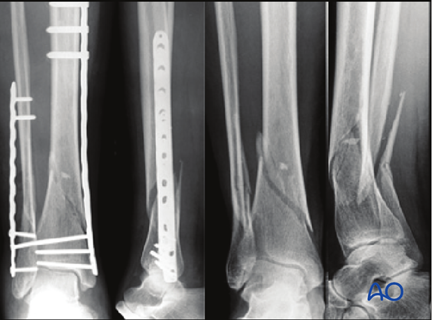
Tibial shaft alignment is influenced by the fibular reduction. If the fibula is too short, a valgus malalignment is likely. This is because the two bones are connected by the syndesmotic ligaments which are usually intact. This problem is demonstrated by the second case, below.
To avoid lateral shortening and valgus deformity, a comminuted fibula is better repaired after the tibia.

4. Pitfalls - Malreduction
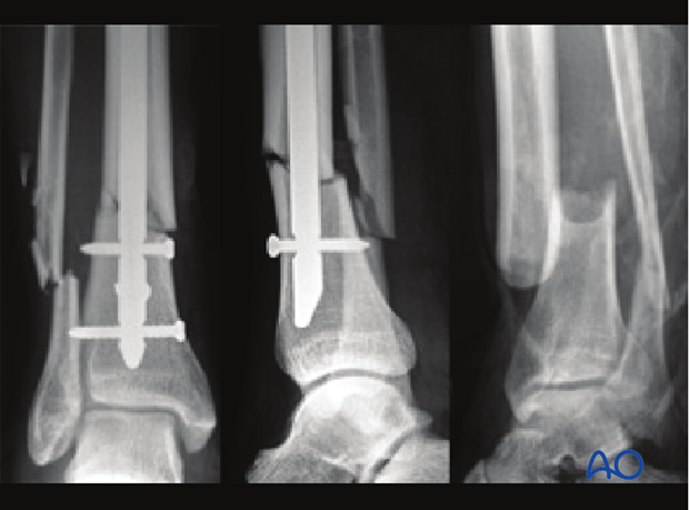
Pitfall flexion-extension malalignment
In this illustrated case, the distal tibia is angulated 10° degrees posteriorly (apex anterior), since the nail is too anterior in the distal fragment.

Pitfall varus-valgus malalignment
In this illustrated case, the ankle joint is malaligned in both planes:
- 10° of valgus due to a unsatisfactory bending of the plate of the tibia (undercontoured). Also the fibula is too short.
- 5° of extension (recurvatum) due to a unsatisfactory indirect reduction of the distal tibia.
The preoperative x-rays are shown on the right.


