Posteromedial approach to the distal tibia
1. Introduction
The posteromedial exposure allows direct reduction of posterior and medial fracture fragments. A posterior plate can be placed, effectively buttressing the posterior fragments. A full thickness subcutaneous anteromedial flap can be created to allow exposure and fixation of the medial malleolus if necessary.
This approach allows for directly buttressing the posterior fracture fragments and allows a second anteromedial incision if necessary.
2. Incision
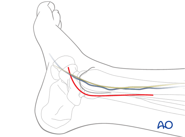
The incision is centered at the ankle joint, between the Achilles tendon and the posteromedial border of the distal tibia. Proximally the incision is parallel to the posteromedial border of the tibia. Distally the incision is parallel to the path of the posterior tibial tendon.

3. Superficial surgical dissection
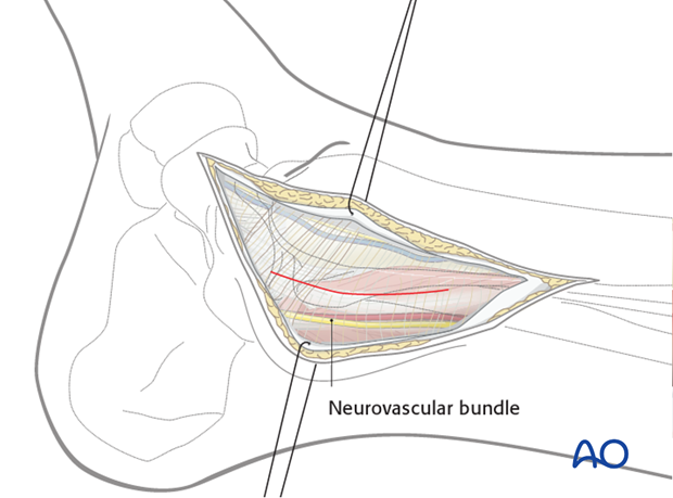
The incision is deepened through the subcutaneous fat and fascia and the deep fascia is revealed over the tendons of tibialis posterior and flexor digitorum longus, the posterior tibial neurovascular bundle and the flexor hallucis longus tendon.
For access to the posteromedial quadrant of the distal tibia, it is necessary to carefully incise the deep fascia proximally, protecting the neurovascular bundle.

4. Deep dissection
The interval used for deep dissection is dependent on the location of the major fracture fragments. It may be located:
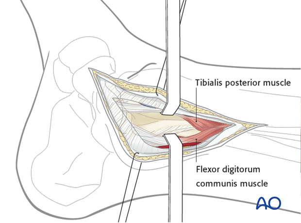
1) Between the tibia and the posterior tibial tendon. This is only useful for proximal exposure as the distal posterior tibial tendon should not be dissected from the posterior tibia.
2) Between the posterior tibial tendon and the flexor digitorum communis (see illustration).

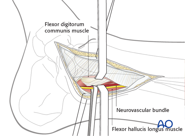
3) Between the flexor digitorum communis and the flexor hallucis longus. This interval requires direct exposure and protection of the neurovascular bundle along its length. The neurovascular bundle can be retracted anteromedially or posterolaterally.

5. Pearls
The posteromedial exposure allows direct reduction of posterior and medial fracture fragments.
A posterior plate can be placed, effectively buttressing the posterior fragments.
A full thickness subcutaneous anteromedial flap can be created to allow exposure and fixation of the medial malleolus if necessary.
Multiple deep surgical intervals can be used dependent on the fracture configuration.


