Minimally invasive approach to the distal tibia
1. Introduction
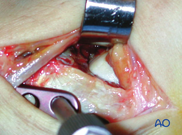
The key concept of this approach is to preserve the soft-tissues and blood supply in the metaphyseal fracture area by not exposing them surgically. An entry site is developed over the distal tibia. The plate is inserted from distal to proximal, through a tunnel between periosteum and intact overlying tissue.
The standard approach for the MIO technique is medially. However, in selected cases with soft-tissue lesions on the medial side, an anterolateral approach can be used.
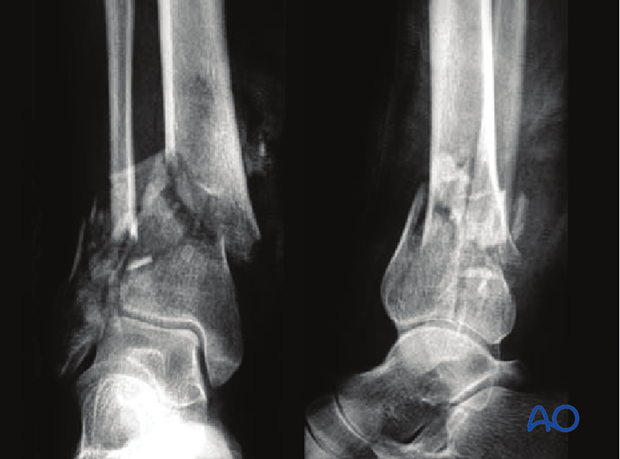
This MIO approach is used for extraarticular fractures, or for simple, minimally displaced, complete articular fractures. In the latter, the articular fracture component is not exposed, and is reduced either by indirect maneuvers using ligamentotaxis, or by the application of percutaneous reduction forceps, or directly by the percutaneously inserted lag screws.
Teaching video
AO teaching video: MIO Tibia, Distal
Medial Approach
2. Skin incision
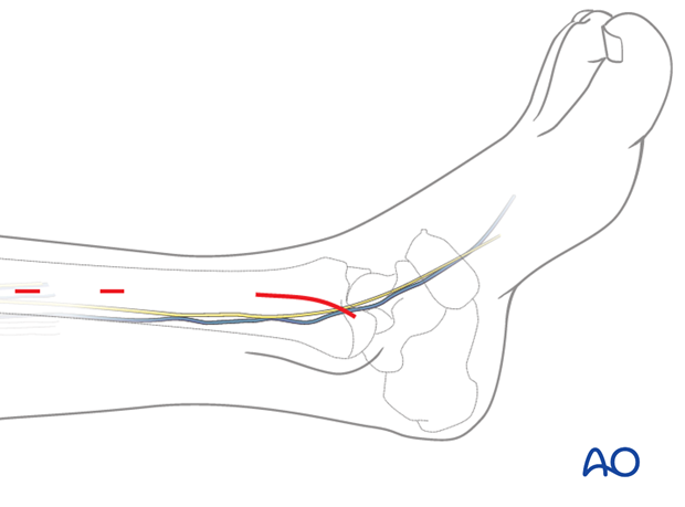
A straight, or slightly curved skin incision is performed on the medial aspect of the distal tibia. The length of the incision varies from 3-5 cm, depending on the type of the planned plate. The incision stops distally at the tip of the medial malleolus.
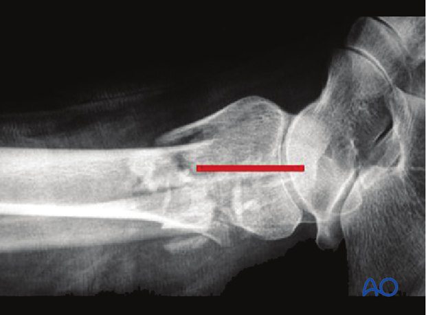
For the insertion of the proximal screws in the diaphysis, separate stab incisions usually are sufficient.
3. Surgical dissection
The incision is carried straight across the subcutaneous fat, preserving the greater saphenous vein and saphenous nerve. They should be held anteriorly with a blunt retractor.
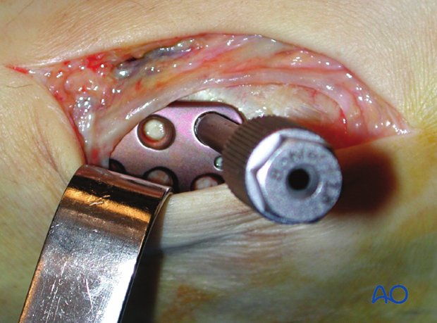
The dissection is advanced down onto the periosteum which is completely preserved. In this anatomical space (epiperiosteal), the tunneling towards the diaphysis can usually easily be achieved with the blunt tip of the plate.
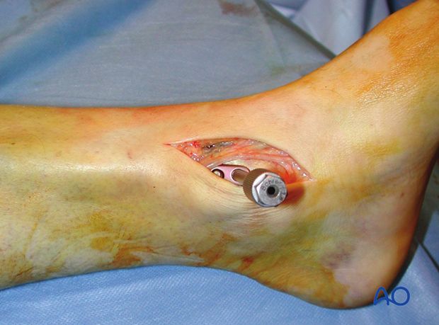
4. Pearl – mini arthrotomy
In case of a fracture extension to the medial malleolus, a small arthrotomy over the medial ankle joint aids removal of intraarticular osteochondral bone debris, inspection of the cartilage surface of the talar dome, and for the visual assessment of the reduction of the medial malleolus.
