Anteromedial approach to the distal tibia
1. Introduction
The anteromedial approach is useful in many types of fractures involving the articular surface, especially if the medial malleolus is also involved.
It is a safe procedure if the correct timing is respected, usually 5-10 days after initial trauma. The skin has to wrinkle, indicating the correct time for surgery.
To prevent postoperative skin necrosis, it is important not to undermine the skin bridge between medial and any lateral approach, and to avoid violation of the anterior tibial tendon sheath.
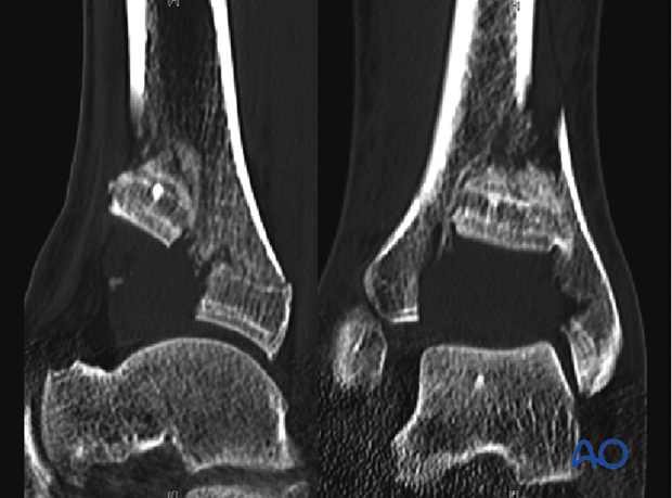
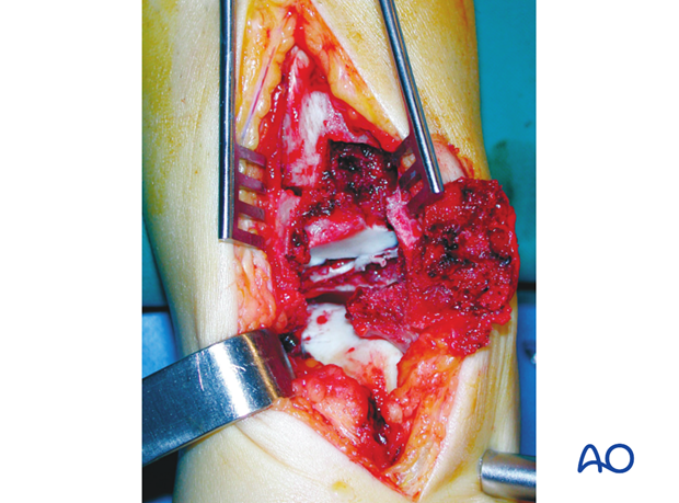
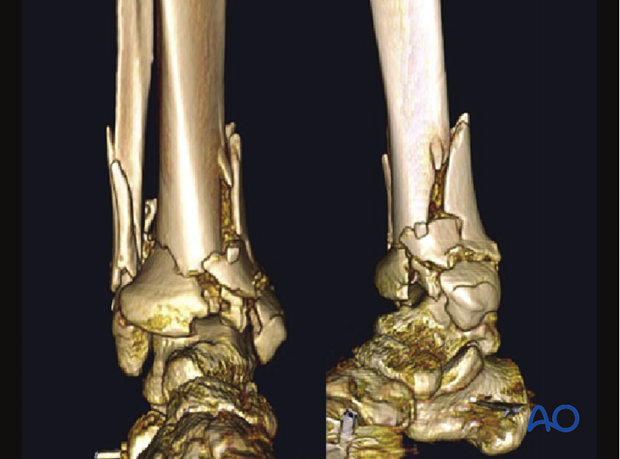
The anteromedial approach has the advantage of excellent visualization of the articular surface in the medial and central part, including the entire medial malleolus. To get access to the anterolateral fragment (Tillaux-Chaput), a small, separate, anterolateral incision might be necessary.
This approach is used for open reduction and internal fixation of the articular part of the tibia. It facilitates accurate articular reduction combined with submuscular and subcutaneous plate applications. It is often used to insert the plate from distal to proximal for bridging the metaphyseal fracture area (combination of limited ORIF and MIO). With the patient in supine position, proximal extension of the incision is unlimited, but usually not required.
2. Skin incision (anterior or medial comminution, planning anterior or medial plate)
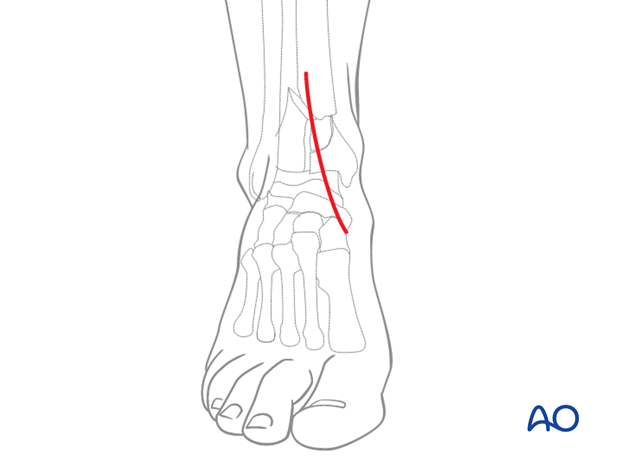
The incision for the anteromedial approach starts about 5–8 cm proximal to the ankle joint just lateral to the palpable tibial crest. It runs in a straight line over the ankle joint towards the base of the navicular, following the medial border of the anterior tibial tendon. A straight incision provides a better approach to the anterior part of the tibia than a curved incision.
3. Surgical dissection
The dissection is deepened through the periosteum, just medial to the anterior tibial tendon. It is critical to leave the tendon sheath intact, and to immediately repair any traumatic or inadvertent disruption that exposes the tendon directly. A medial plate can be slid in a MIO fashion.
Minimal exposure and careful handling of the periosteum are essential to prevent any further vascular damage of the fracture fragments.
Lateral dissection between the posterior border of the tendon sheath and the periosteum is performed to get access to reduce the anterolateral fragment. However, for fixation (screw insertion) it might be necessary to have a separate small anterolateral incision.
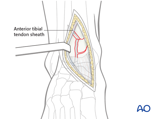
The tibiotalar joint is opened in the sagittal direction, usually in line with the fracture line between the two main anterior articular fragments.
Any transverse incision of the anterior capsule to further expose the joint should be kept short as this risks devascularization of the anterior fragments (supplied by branches of the anterior tibial artery).
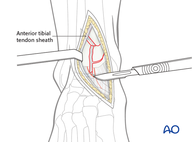
4. Exposure of the joint
A large distractor, from tibia to medial talus, pulls the talus distally, aiding exposure. A bone spreader can be used to separate the anteromedial and the anterolateral articular fragments. This exposes the joint, allowing an excellent approach to the center as well as to the posterior part of the fracture.
