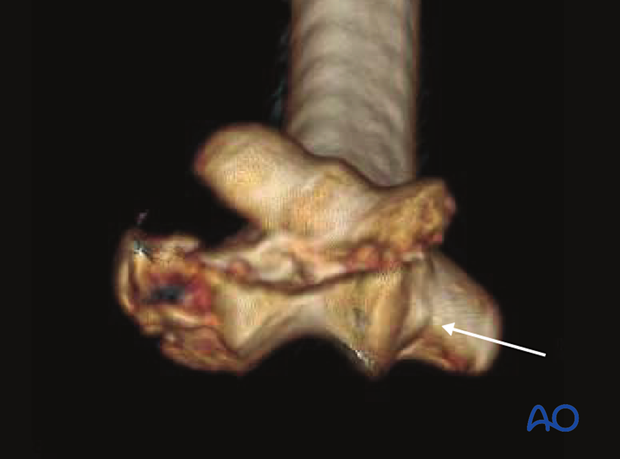
13B3 Partial articular fracture in frontal/coronal plane
Introduction
Partial articular fractures in the frontal/coronal plane are classified by the AO/OTA as 13B3.
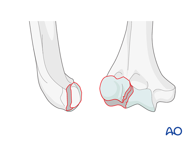
This fracture group addresses coronal plane fractures of the capitellum and part of the trochlea without fracture of the lateral column.
Capitellar fracture
Definition
This fracture type is classified by the AO/OTA as 13B3.1.
Further characteristics
Take care when assessing an apparently simple capitellar fracture. These are often complex. Many extend into the lateral trochlea.
Many fractures also involve a fracture of the lateral epicondyle and impaction of the posterior aspect of the lateral column or the trochlea.
Computed tomography with 3D reconstruction is necessary to identify the fracture characteristics and facilitates planning.
X-ray
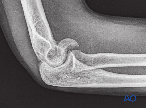
Lateral view showing anterior displacement of the capitellar fragment
3-D CT
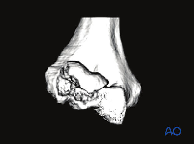
This CT image of the same case shows extension of the fracture into the lateral aspect of the trochlea. This extension is difficult to diagnose on plain x-rays. Therefore, CT imaging of articular fractures facilitates the diagnosis and planning of surgery.
Trochlear fracture
Definition
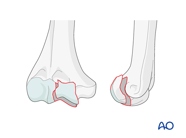
This fracture type is classified by the AO/OTA as 13B3.2.
Further characteristics
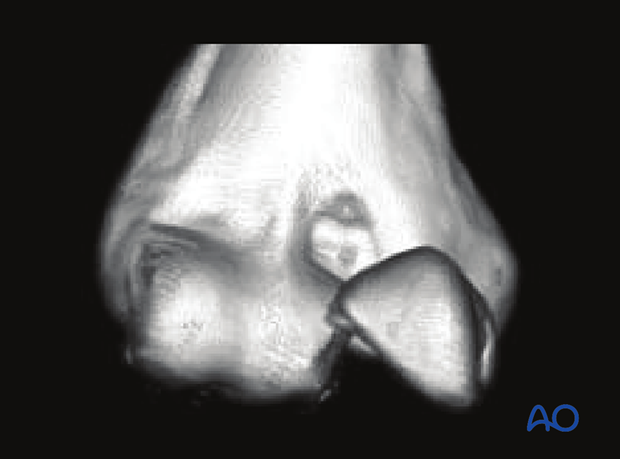
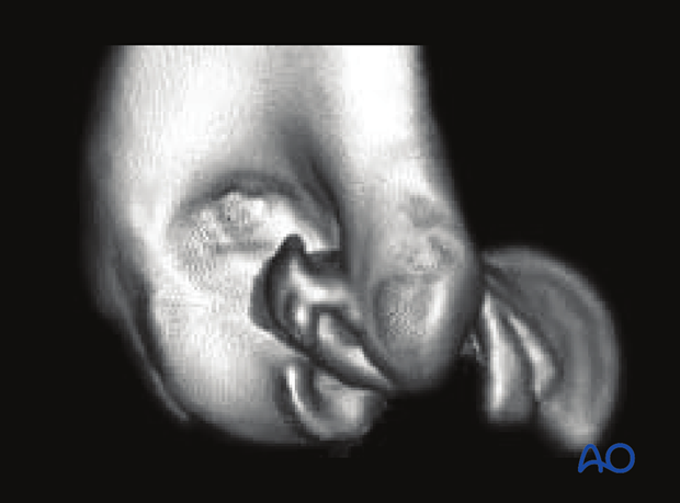
Trochlear fractures are difficult to see on x-rays. Computed tomography—with 3-D reconstruction—is particularly useful for understanding the fracture anatomy.
There is often complex articular comminution.
3-D CT
AP view of anteriorly displaced partial trochlear fragment
Lateral view of the same case
Capitellar and trochlear fracture
Definition
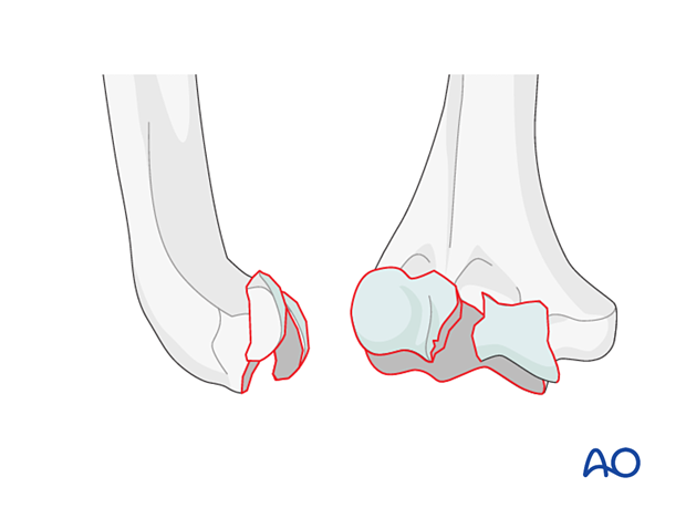
This fracture type is classified by the AO/OTA as 13B3.3.
Further characteristics
These fractures are often associated with impaction of the posterior part of the lateral column and/or the posterior trochlea.
Most apparently isolated capitellar fractures are more complex than they initially appear.
Computed tomography—particularly 3-D CT—can help to define the fracture anatomy and facilitate the planning of the surgery.
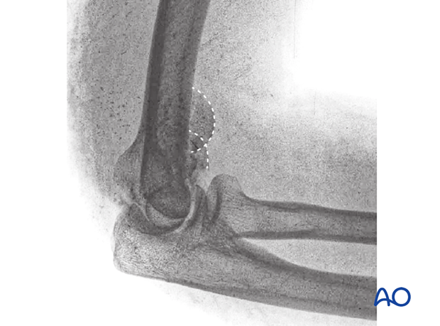
X-ray
The ‘double arc’ sign is associated with displacement of a complete coronal fracture of the combined capitellum and trochlea.
3-D CT
This is the CT scan of the above case. Computed tomography, with 3-dimensional reconstruction, in particular, is very useful for understanding apparently simple capitellar fractures that turn out to be more complex.
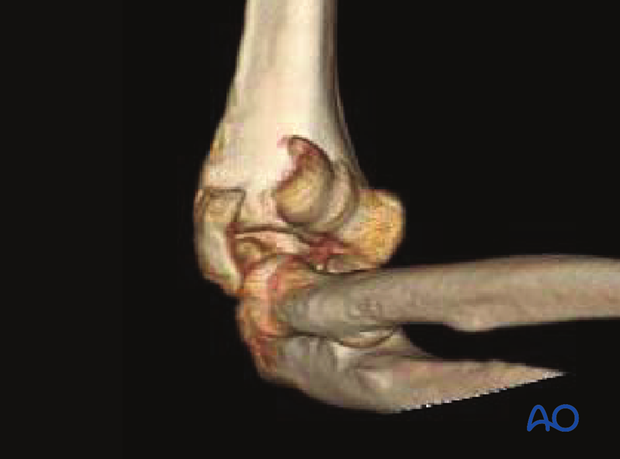
In this image, it is apparent that the articular fragments may not correctly align, suggesting impaction of the posterior trochlea and particularly the lateral column.
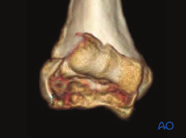
An end-on view shows the complexity, particularly on the lateral side.
Also, one can see a fracture—albeit very subtle—between the medial epicondyle and the medial trochlea.
The surgical exposure should take this into account.
