Posterior triceps-split approach (Campbell) to the distal humerus
1. Introduction
The main advantage of this approach is its extensile nature and that it avoids extensive dissection of the ulnar nerve.
The main disadvantage is that it provides limited exposure to the articular surface. The triceps insertion may be elevated from the olecranon but must be repaired to avoid triceps insufficiency.
2. Skin incision
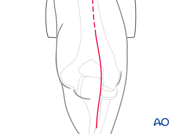
Make an incision centered on the junction of the middle and distal thirds of the humeral shaft. Avoid placing the incision over the tip of the olecranon. Some surgeons make a straight incision slightly medial or lateral, whereas others prefer a curved incision. The incision ends over the ulnar diaphysis.
Elevate full-thickness fasciocutaneous flaps to protect the cutaneous nerves.
3. Split of the triceps tendon
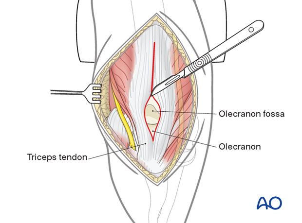
Dissect the subcutaneous tissue from the deep fascia.
Split the triceps tendon in the midline from the tip of the olecranon to the upper limit of the olecranon fossa.
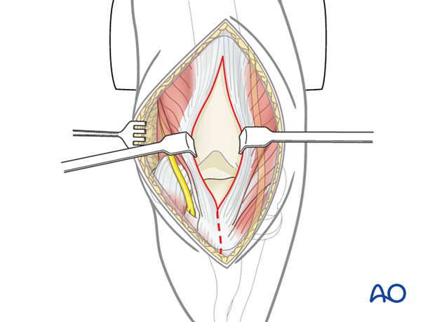
Insert two smooth soft-tissue retractors.
Further split the triceps tendon and aponeurosis proximally (normally 3–4 cm proximal to the fracture line). If a more proximal exploration of the distal humerus is required, the triceps muscle may be split further, but the radial nerve is at risk in the proximal aspect and should be identified.
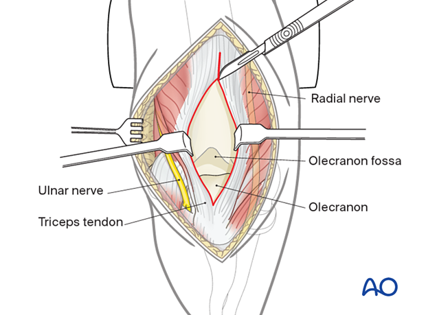
Subperiosteal dissection of the tendon and associated soft-tissue attachments of the ulna should be performed carefully.

Soft-tissue retraction will now provide a wide view of the posterior aspect of the distal humerus.
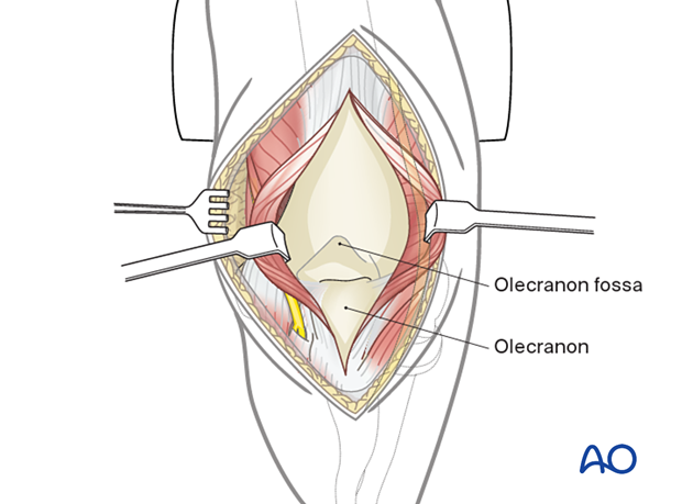
4. Wound closure
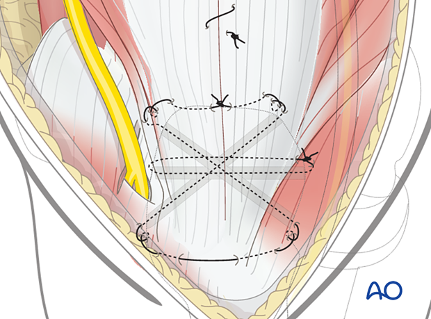
The muscle split can be repaired with a continuous absorbable suture.
Distally, diligent repair of the triceps insertion to the olecranon is required. It is recommended to use transosseous sutures to achieve this.
Prepare transverse and oblique transosseous tunnels in the olecranon using a 2–2.5 mm drill bit.
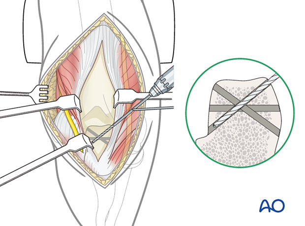
Oppose the tendon firmly to the olecranon using nonabsorbable sutures passed through these tunnels and the tendon with a straight needle.
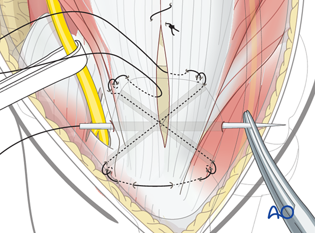
The patient should avoid resisted extension activities while the tendon heals to avoid triceps insufficiency.
