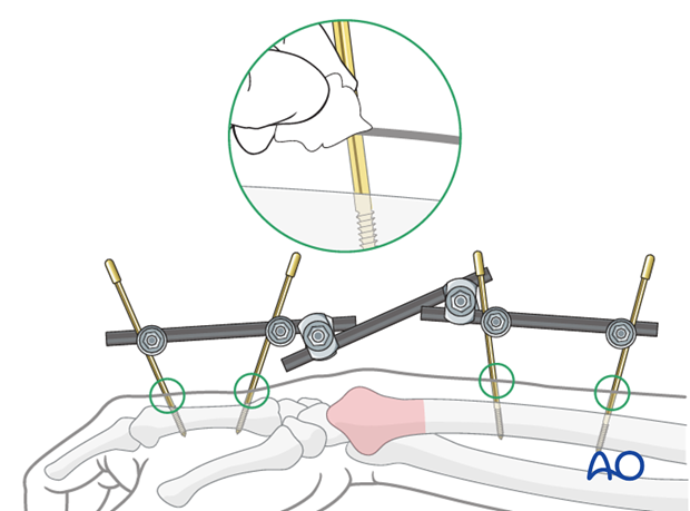
Closed reduction - K-wires and cast/external fixator
1. Preliminary remarks
Fracture assessment
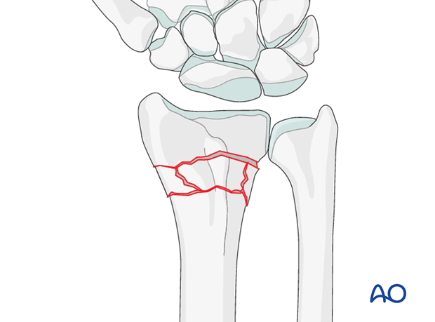
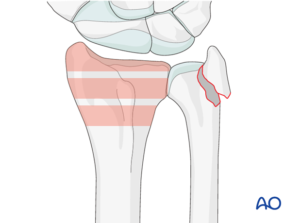
These fractures are extraarticular but multifragmentary. They may often be associated with an ulnar styloid fracture.
Indications
Because of the comminution, these fractures are usually treated with ORIF. In patients not suitable, a simple cast is likely to be inadequate, but the addition of K-wires may provide some degree of stabilization.
Anatomy of the wrist
A thorough knowledge of the anatomy around the wrist is essential. Read more about the anatomy of the distal forearm.
2. Associated injuries
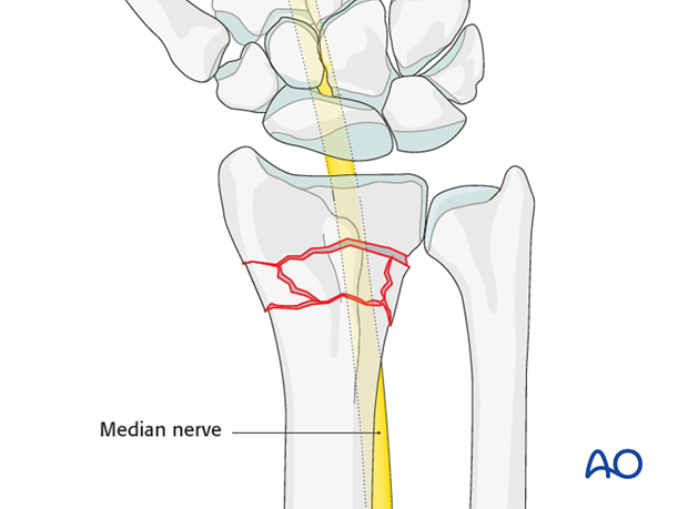
Median nerve decompression
If there is dense sensory loss, or other signs of median nerve compression, the median nerve should be decompressed.
Associated carpal injuries
These injuries may be associated with shearing injuries of the articular cartilage, scaphoid fracture and rupture of the scapholunate ligament (SL). Every patient should be assessed for this injury. If present, see carpal bones of the Hand module.
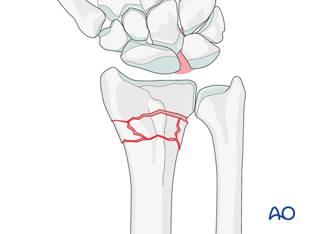
DRUJ/ ulnar injuries
These injuries may be accompanied by avulsion of the ulnar styloid and/or disruption of the DRUJ. If there is gross instability after the fixation of the radial fracture, it is recommended that the styloid and/or the triangular fibrocartilaginous disc (TFC) is reattached. This is not common in simple fractures, but may occur with some high energy injuries.
3. Patient preparation
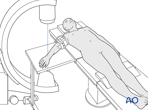
The patient may be placed either in a supine position for palmar approaches or for dorsal approaches.
4. Closed reduction
Traction
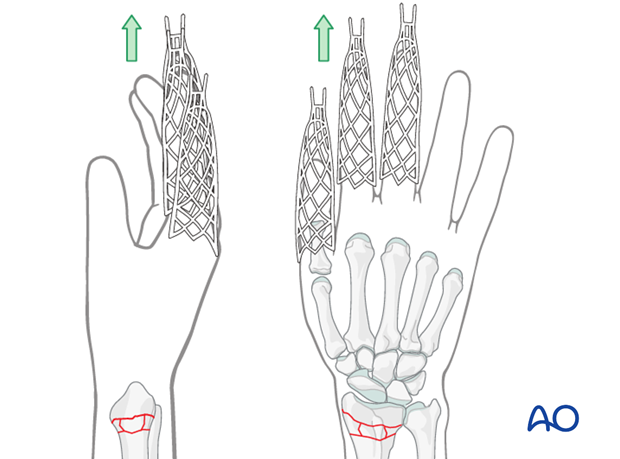
Closed reduction can be performed with or without continuous finger traction, e.g., using Chinese finger traps.
Closed reduction of extraarticular multifragmentary fractures
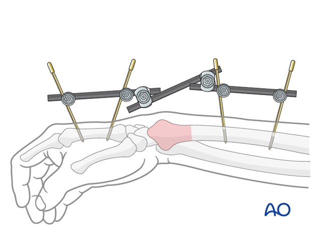
Longitudinal traction is essential to restore radial length, but attention also has to be paid to correcting any associated angular deformity - dorsal, palmar and/or radial. In very unstable fractures, reduction may be achieved with a temporary spanning external fixator. This will hold the reduction whilst the stabilizing K-wires are inserted, and in many cases, it can then be removed.
5. K-wire insertion
Preliminary remark
There are numerous techniques of K-wire fixation (e.g., two wires, three wires, Kapandji-technique) for fractures of the distal radius.
We describe a technique using three K-wires. Two are introduced from the tip of the radial styloid, one from the dorsoulnar aspect.
Insertion of the first smooth K-wire
First, a 1 cm incision is made over the tip of the radial styloid. The radial styloid is exposed by blunt dissection and great care is taken not to injure the superficial branch of the radial nerve or the tendons of the first and third extensor compartments. The drill guide is introduced between the tips of the soft-tissue spreader.
After checking reduction and anticipated direction of the K-wire using image intensification, the K-wire is introduced carefully with a power drill.
The K-wire should just penetrate the opposite cortex of the radial shaft.
When inserting the first K-wire, additional control of the distal fragment may be necessary. This is best achieved by using a small pointed awl, inserted percutaneously.
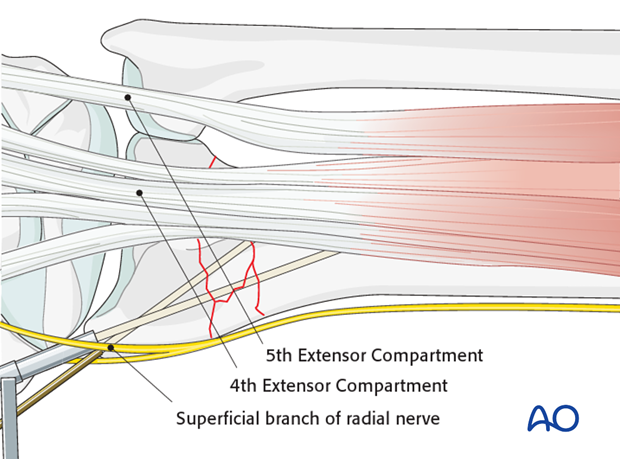
Second K-wire
A second K-wire is introduced through the radial styloid in the same manner, but in a divergent direction.
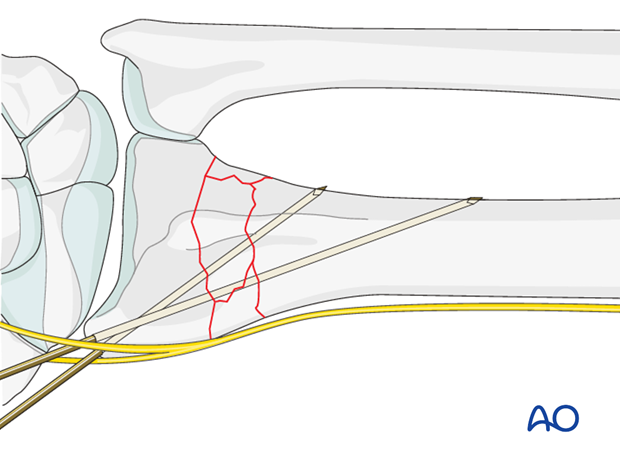
Third K-wire: Insertion from the dorsoulnar aspect
A second incision is made between the fourth and fifth extensor compartments. Blunt dissection to the bone is carried out.
Under image intensifier control, the third K-wire is introduced from the dorsoulnar rim of the radius into the anterior cortex of the radial shaft.
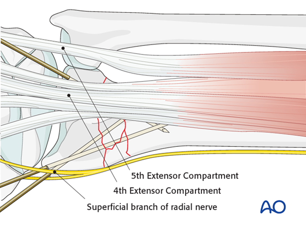
The fourth compartment is displaced radially by the pressure of the thumb, which enables precise K-wire positioning into the dorsoulnar corner of the lunate facet.
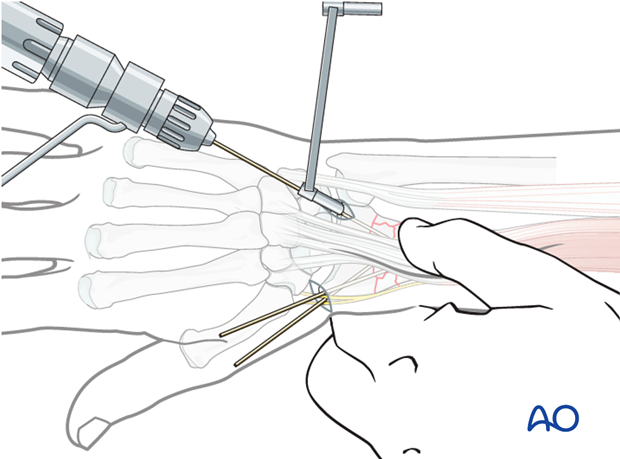
Cut and bend K-wires
The ends of the wires should be cut and bent.
The ends may be left underneath the skin, to reduce the possibility of pin-track infection.
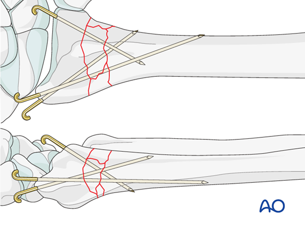
Pitfall: K-wire crossing
The K-Wires should not cross at one point at the fracture level.

Pearl: Alternative method
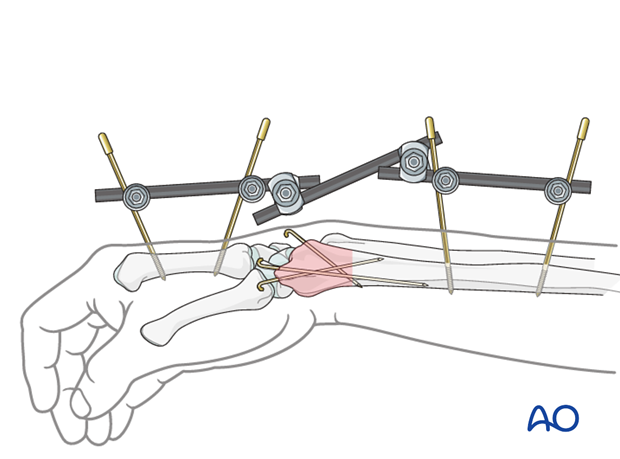
If there are concerns about the security of reduction in a cast, particularly a risk of shortening, an external fixator as neutralizing device (without traction) may be preferred after K-wire fixation.
This is particularly useful in extensive metaphyseal comminution or osteoporosis.
6. Cast application
For more details on casting techniques, see nonoperative treatment options.
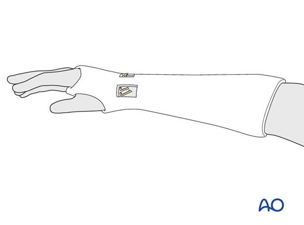
A well-padded cast is applied.
One option to consider is creating windows in the cast directly over the pin sites.
Because the reduction is stabilized with K-wires, a below elbow cast is preferred, and molding is less important.
7. Aftercare
Functional exercises
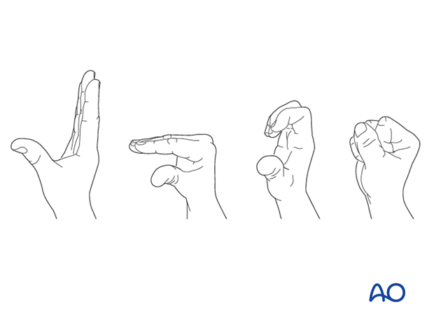
As soon as possible, the patient should be encouraged to elevate the limb and mobilize the digits, elbow and shoulder.

If necessary, functional exercises can be under the supervision of a hand therapist.
Follow up
See patient 7-10 days after surgery for a wound check and/or suture removal. X-rays are taken to check the reduction. Refer to your local protocol for timing of X-ray follow-up.
K wire removal
The K-wires are usually removed at about 6-8 weeks. If the wires were buried, it may be necessary to take the patient to the OR to reopen the incisions and retrieve the wires.
Cast removal
The cast is left in place for 4-6 weeks. An x-ray will document fracture position at this time.
Exfix management
The patient needs to be instructed in pin care and should clean the insertion sites daily.
The external fixator is usually left in place for six weeks, but in very unstable fractures or where there are delays in fracture healing it may be left for longer. Excessively long application of an external fixator risks joint stiffness.
The timing of external fixator removal is influenced by various factors. These include the specific details of the fracture and patient, and the radiological appearance of the healing fracture.
