ORIF - Joint-spanning distraction plate
1. Preliminary remarks
Fracture assessment
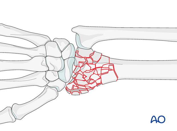
As these are intraarticular fractures, where possible, they should be treated with anatomic reduction and absolute stability in order to minimize the risk of subsequent degenerative changes in the joint.
Anatomical reduction and stabilization of these articular fractures is also essential because of the functional implications of the involvement of the distal radioulnar joint.
Complete articular fractures are among the most common fractures seen and treated in the older population, with underlying osteoporosis. When these fractures occur in younger individuals, they are more likely to be the result of high energy trauma, with associated soft-tissue, or skeletal injuries.
It is not possible to make an accurate assessment of the details of these injuries without a CT scan.
In cases of extreme comminution, where it is not felt possible to reconstruct the joint, a joint spanning bridge plate of the distal radial joint surface may be considered.
Provisional reduction
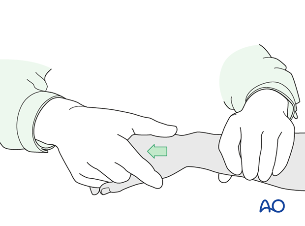
Reduction is achieved by applying longitudinal traction either manually or using Chinese finger traps.
The reduction is maintained by a temporary splint.
If definitive surgery is planned, but cannot be performed within a reasonable time scale, a temporary external fixator may be helpful.
2. Associated injuries
Median nerve compression
If there is dense sensory loss, or other signs of median nerve compression, the median nerve should be decompressed.

Associated carpal injuries
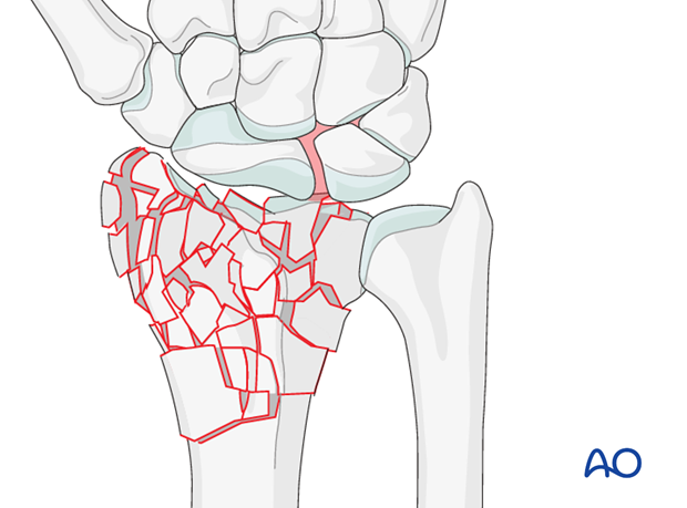
These injuries may be associated with shearing injuries of the articular cartilage, scaphoid fracture and rupture of the scapholunate ligament (SL). Every patient should be assessed for this injury. If present, see carpal bones of the Hand module.
DRUJ/ulnar injuries
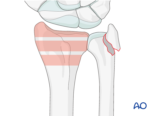
These injuries may be accompanied by avulsion of the ulnar styloid and/or disruption of the DRUJ. If there is gross instability after the fixation of the radial fracture, it is recommended that the styloid and/or the triangular fibrocartilaginous disc (TFC) is reattached. This is not common in simple fractures, but may occur with some high energy injuries.
The uninjured side should be tested as a reference for the injured side.
It may not be possible to assess DRUJ stability until the fracture has been stabilized (as described below).
3. Patient preparation and approach
Patient preparation
This procedure is normally performed with the patient in a supine position for dorsal approaches.
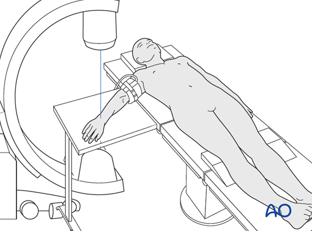
Approach
For this procedure an extended dorsal approach is normally used.
A thorough knowledge of the anatomy around the wrist is essential. Read more about the anatomy of the distal forearm.
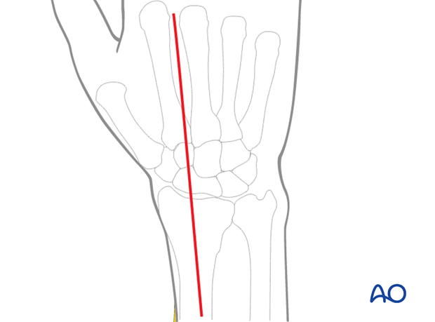
4. Open reduction
The main fragments are reduced to reconstruct the distal radial joint surface as accurately as reasonably possible. The proximal carpal bones may be used as a template against which the fragments are reduced. A dental pick, K-wires or a small periosteal elevator are used for the reduction.
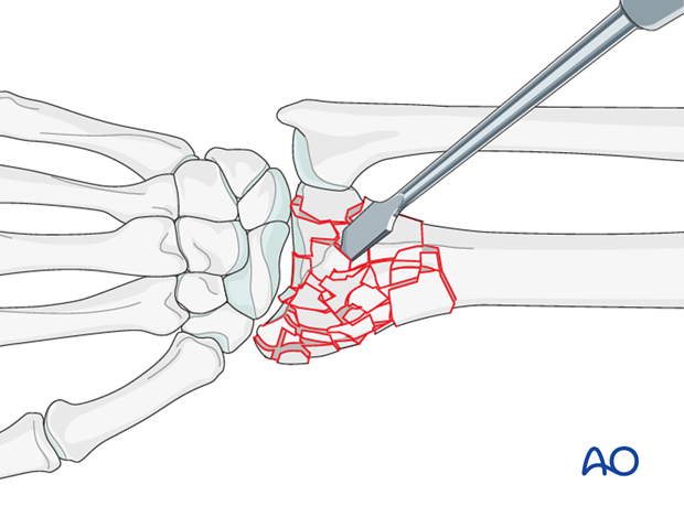
If there are fragments large enough to be secured with a K-wire, these may be used for limited fixation of the fracture.
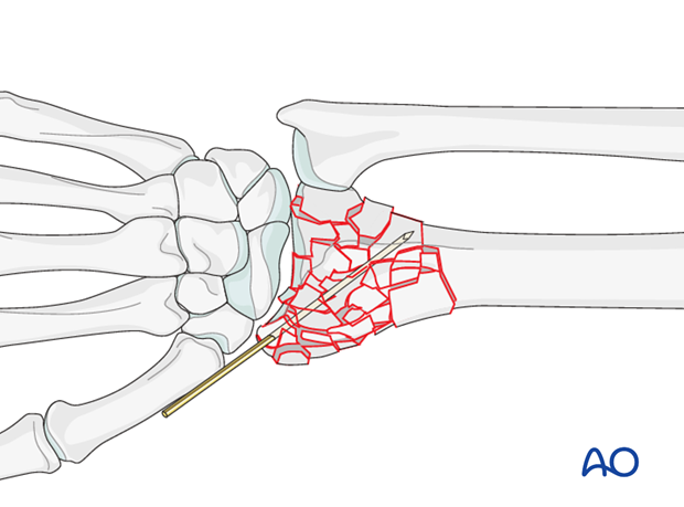
5. Plate application
Plate
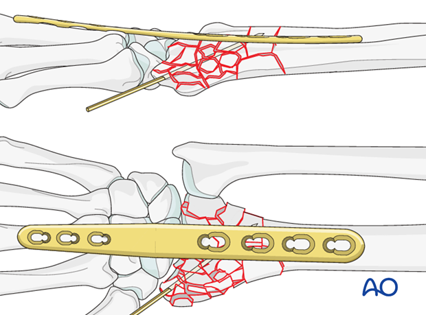
A straight wrist fusion plate or a standard 3.5 plate is contoured and slightly bent to fit the dorsal side of the distal radius, the carpus and the third metacarpal, holding the wrist in slight extension.
The aim is to hold the carpus in the correct relationship to the radius for the multiple fragments to heal in a reasonable approximation to an anatomic position.
Screw insertion
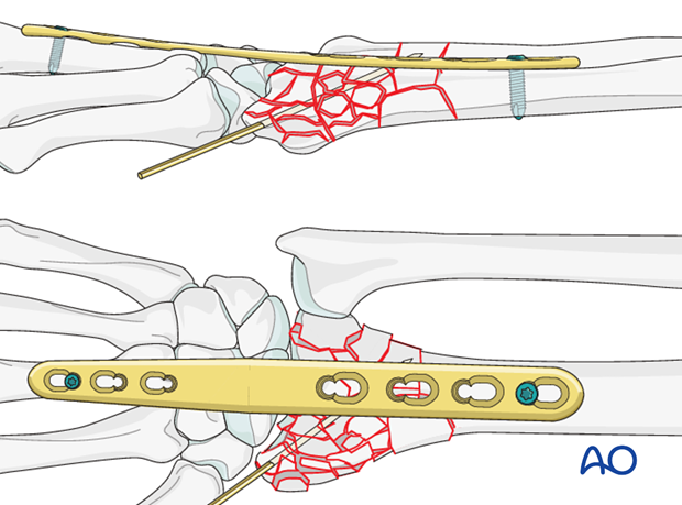
The plate is first fixed on the (third) metacarpal with a screw in the most distal plate hole.
The plate is aligned on the distal radius, with no more than 5 mm distraction at the radiocarpal joint, and held with a fracture reduction clamp.
The forearm is supinated approximately 30° and the reduction assessed.
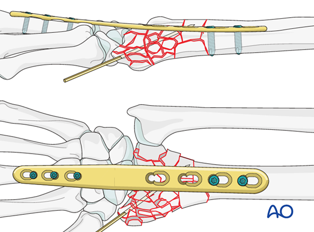
If the fracture position and forearm rotations are satisfactory, three proximal screws are inserted, and two further screw are inserted into the radial shaft.
If a locking plate is applied, locking head screws should be used.
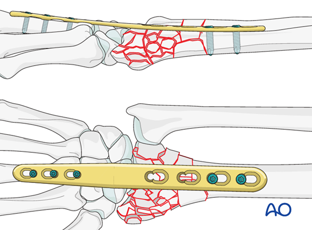
The K wires may be removed at approximately six weeks, but the plate is left in place until bone healing has been confirmed radiologically, usually between three to four months.
It is then removed an active rehabilitation program is initiated.
6. Aftercare
Functional exercises
Immediately postoperatively, the patient should be encouraged to elevate the limb and mobilize the digits, elbow and shoulder.
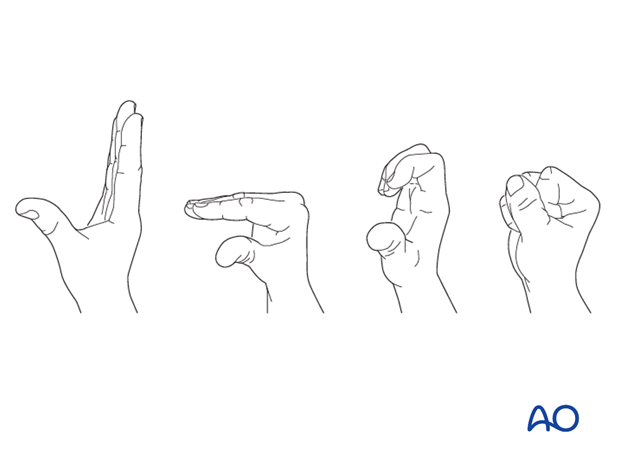
Some surgeons may prefer to immobilize the wrist for 7-10 days before starting active wrist and forearm motion. In those patients, the wrist will remain in the dressing applied at the time of surgery.

Wrist and forearm motion can be initiated when the patient is comfortable and there is no need for immobilization of the wrist after suture removal.
Resisted exercises can be started about 6 weeks after surgery depending on the radiographic appearance.
If necessary, functional exercises can be under the supervision of a hand therapist.

Follow up
See patient 7-10 days after surgery for a wound check and suture removal. X-rays are taken to check the reduction.
Implant removal
Implant removal is purely elective but may be needed in cases of soft-tissue irritation, especially tendon irritation to prevent late rupture. This is particularly a problem with dorsal or radial plates. These plates should be removed between nine and twelve months.


