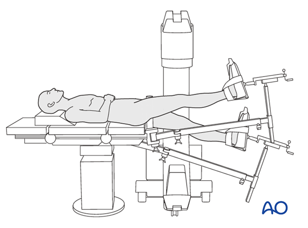
Scissor position
1. Introduction
Scissoring makes length, alignment and rotational confirmation easy. Raising the injured leg facilitates reduction of any flexed proximal fragment (iliopsoas muscle).
Careful pre-cleaning of the soft tissues should be performed especially if gross contamination occurs.

2. Preoperative preparation
Operating room personnel (ORP) need to know and confirm:
- Site and side of the fracture
- Type of operation planned
- Ensure that the operative site has been marked by the surgeon
- Condition of the soft tissues (fracture: open or closed)
- Implant to be used
- Patient positioning
- Details of the patient (including a signed consent form and appropriate antibiotic and thromboprophylaxis)
- Comorbidities, including allergies
Note: Improve the preoperative preparation with the WHO Surgical Safety Checklist.
3. Anesthesia
This procedure is performed with the patient under general or regional anesthesia
Long-lasting postoperative complete pain blocks for the patient with injured leg should be avoided as this could hide symptoms of a subsequent compartment syndrome.
4. Positioning
- Supine with bilateral boots and traction
- Reconfigure the fracture table to establish the bilateral traction boot position and transfer the patient to a fracture table.
- Position the fractured leg with traction in a 20° hip flex position with traction. The unaffected leg is positioned in a 30° hip extension position on the other side of the post in a traction boot.
- Reduce the fracture with traction and manipulation before preparing and draping the patient.
- Pad all pressure points carefully (especially in the elderly). Place the ipsilateral arm across the chest to be out of way.
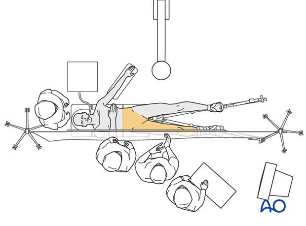
- Position the image intensifier on the opposite side of the injury and perpendicular to the patient.
- Ensure that you can get good-quality AP and lateral x-ray views of the entry point (piriform fossa should be more easily reached with the affected leg slightly adducted), fracture site, and distal femur before draping.

5. Skin disinfecting and draping
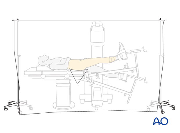
- Maintain traction on the limb during preparation to avoid excessive deformity at the fracture site.
- Disinfect the exposed area from above the iliac crest to the mid-tibia with the appropriate antiseptic. Free drape the affected limb or use a vertical isolation drape.
- Ensure the adhesive portion of the drape is large enough to reach from the iliac crest to the knee joint to allow distal locking.
- A single-use exclusion drape is used.
- Place the image intensifier on the nonsterile side of the exclusion drape.
- Drape the image intensifier.
- Traditional drapes may be used. Ensure a waterproof environment for the operative site.

6. Operating room set-up
- Position the operating table (if feasible) within the operating room to allow maximum space on the operating side for the surgeon, staff, and trolleys.
- The surgeon, assistant, and ORP stand on the side of the injury.
- Place the image intensifier on the opposite side of the patient, perpendicular to the patient.
- Place the image intensifier display screen in full view of the surgical team and the radiographer at the foot of the table.


