Sustentacular fracture of the calcaneus body
General considerations
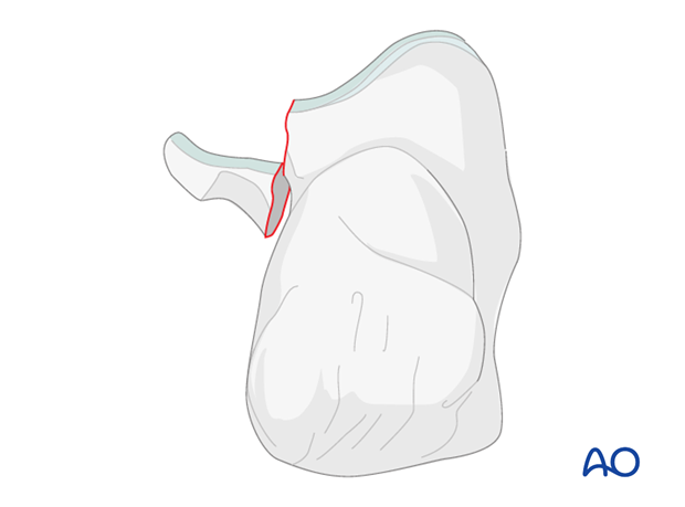
This fracture occurs with a fall from height, usually accompanied by a twisting mechanism. Sometimes this injury is minimized or missed and it is not until continued symptoms push for further investigation that a diagnosis is made.

Radiology
X-rays
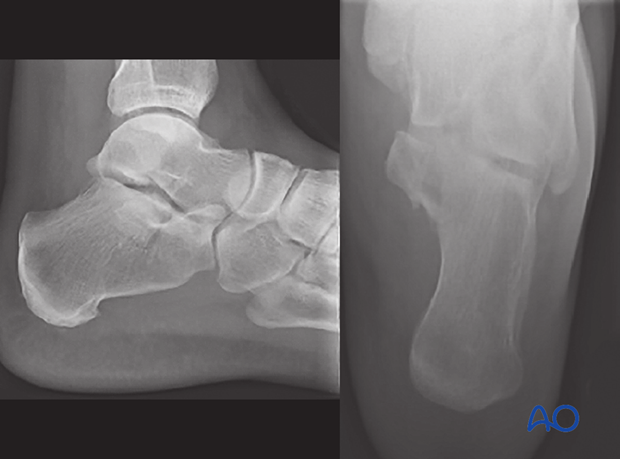
The key to this injury’s diagnosis involves careful and correct x-ray and x-ray interpretation. Lateral x-rays of the hindfoot and axial views are sufficient, but often misinterpreted, or minimized.

CT
Initially, the diagnosis of this fracture is usually missed because it is difficult to pick up on normal x-rays of the foot. Continuing pain often leads to a CT scan. The diagnosis then becomes obvious.
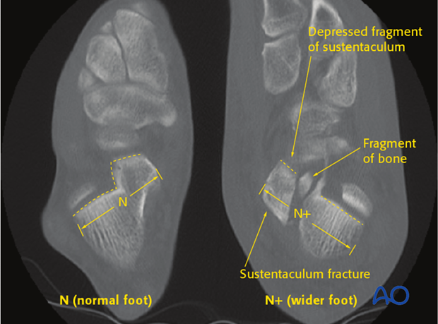
As this injury occurs with axial load, the sustentaculum remains attached to the talus and is depressed plantarwards immediately with the os calcis and the foot migrating laterally and upwards.
As seen with the normal CT on the left, the middle facet should always be more superior and not depressed, as is demonstrated by the CT on the right. Note the tremendous widening of the hindfoot.

Surgical indications
As this injury makes the three facets of the subtalar joint incongruent, most sustentacular fractures, when displaced, require surgery. Only undisplaced fractures and fractures in the very elderly would be treated nonoperatively. Accurate reduction is almost always indicated for good subtalar function.


