Displaced fracture of the calcaneus body
General considerations
Fractures of the calcaneus are common and account for approximately 60% of tarsal injuries. They result from an axial load and may demonstrate varying severity and location of fracture type.
The type of fracture is determined by position of the foot, direction and magnitude of the force and the quality of the bone. Patients will present with pain and swelling, with inability to bear weight.
Bilateral fractures of the calcaneus occur approximately 10% of the time.
Radiology
Radiographic views
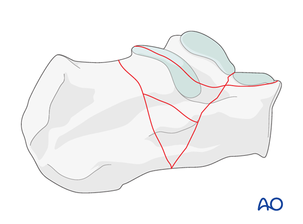
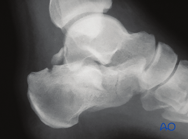
The radiographic assessment begins with 3 views of the foot as well as an axial (Harris) view. The basic fracture types, i.e. tongue type vs joint-depression type, are best demonstrated in the lateral view (as in this image).
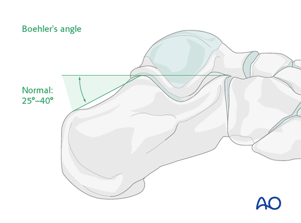
Böhler’s angle
A decrease in Böhler’s angle demonstrates the severity of joint injury and displacement (depression) as measured on the lateral x-ray. The normal angle is 25 degrees to 40 degrees. If this angle remains above 15 degrees, nonoperative care can be suggested.
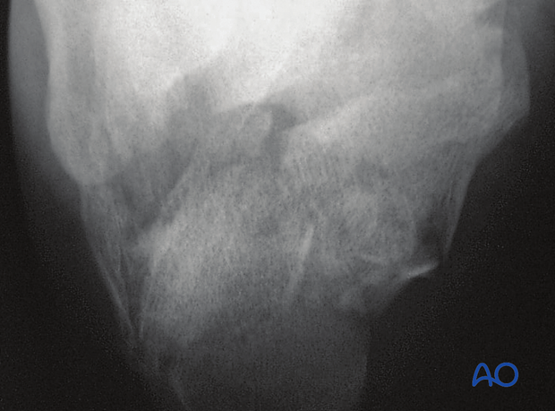
Axial view
The axial view shows the primary joint displacement and angulation of the tuberosity as well as any increase in heel width.
This view is important intra-operatively to ensure that there is no varus in any calcaneal reconstructive procedure.
This is a difficult x-ray to obtain preoperatively.
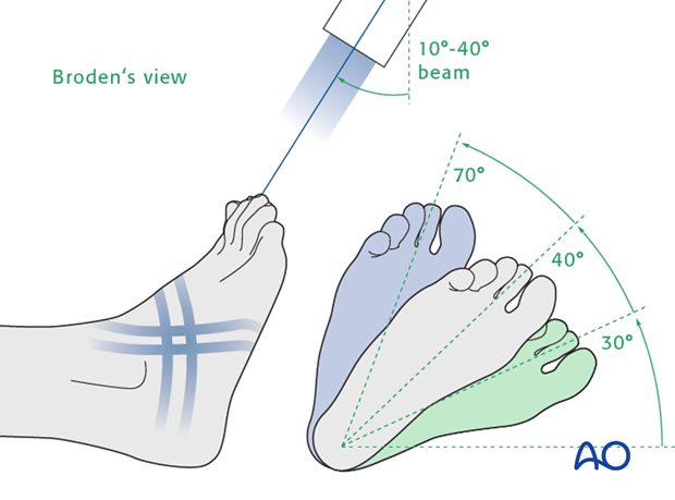
Broden's view
Broden's views are special calcaneal radiographhic projections to show the congruence of the subtalar joint.
They are taken at 30°, 50° and 70° to the horizontal.
Computer-assisted tomography
Computer-assisted tomography is essential for accurate diagnosis and for surgical planning.
Axial CT images demonstrate fracture extension into the calcaneo-cuboid joint and tuberosity.
Coronal CT images depict the involvement of the posterior facet as well as shortening and position of the tuberosity.
Sagittal CT reconstructions can further elucidate the injury.
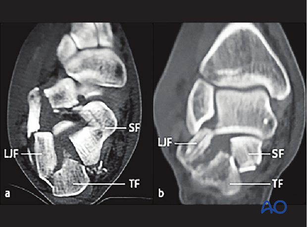
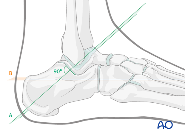
For best CT imaging careful foot position is mandatory.
As shown in the illustration the planes of reconstruction are A perpendicular to the post facet (semi-coronal) and B parallel to the sole of the foot (not metatarsal).
Müller/AO/OTA classification
No single classification encompasses all calcaneal fractures.
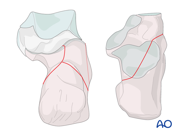
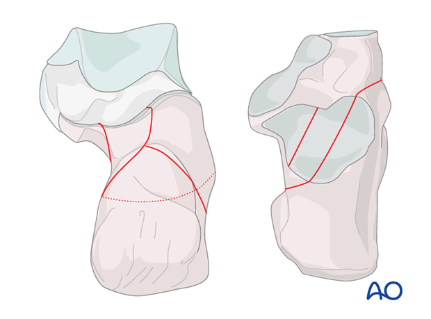
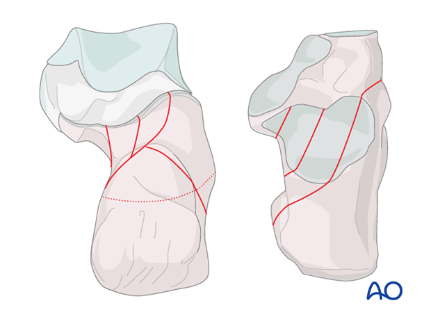
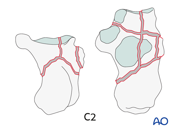
The Müller/AO/OTA classification divides the fractures into varying types, A, B and C. By identifying the fracture type we obtain information as to the severity of the fracture and its likely outcome, which is most helpful in decision making as to the type of treatment.
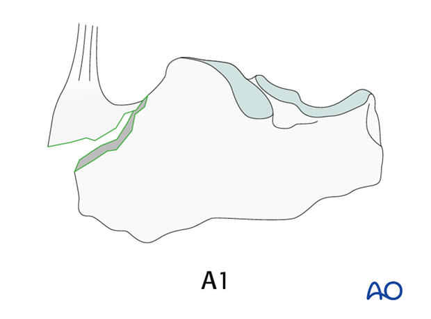
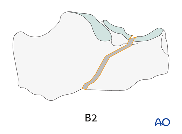
A1 and B2 fractures are examples of extraarticular injuries where surgery is indicated despite the fact that the fracture is extraarticular.
The C2 fracture demonstrates a degree of joint involvement which is significant but still amenable for a successful surgical reconstruction. As the fracture involves the articular surface, an anatomic reduction is a must.
In all of these fracture types the prognosis improves if the anatomy of the os calcis is returned to normal.
Essex-Lopresti classification
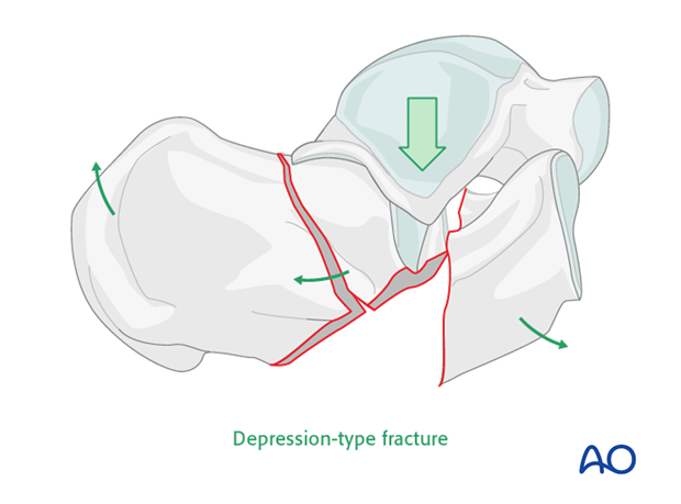
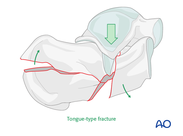
This classification was first published in 1951-52*. It is useful in separating the joint fractures into two types. We see first an example of a “joint depression” and in the lower image a “tongue type”. The joint depression is notorious for the degree of joint involvement and displacement.
* Essex-Lopresti P. The mechanism, reduction technique, and results in fractures of the os calcis, 1951-52. Br J Surg. 1952 Mar;39(157):395-419
Note that in the tongue-type fracture the fracture exits posteriorly, splitting the posterior portion into two. Different fixation techniques are required in order to reduce and stabilize this deformity.
Sanders classification
Sanders based his classification on the CT coronal projection of the articular surface. The prognosis worsens with the increase of articular comminution, and in addition the surgical reconstruction becomes more difficult not only with the comminution, but with the translation of the fracture lines closer to the medial wall.
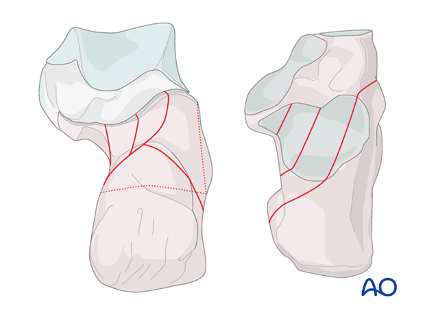
2-part joint injuries are relatively easy to reduce and have a better outcome while the 3- and 4-part displaced intraarticular fractures will be much more challenging, and are increasingly more difficult to reduce. Their outcome is guarded.
