Decision making: choice of exposure
1. Choosing between anterior and posterior approach
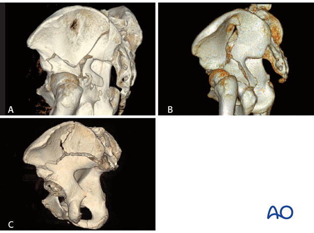
Five of the ten fracture types involve both the anterior and posterior column but usually only either an anterior or posterior approach in isolation is used.
For example, this transverse fracture extends through both the anterior and posterior column.
The surgical approach is dictated by the height of the fracture (transtectal, juxtatectal, infratectal), the fracture obliquity, and the pattern of displacement.
For mainly posterior displaced fractures, the Kocher-Langenbeck will be sufficient exposure to reduce the fracture without a second direct anterior exposure. This is because the displacement is typically greater posteriorly and the retroacetabular surface offers excellent opportunities for both reduction tools and fixation strategies.
Furthermore, reduction of the anterior elements is accomplished by manipulation of the posterior aspect of the ischiopubic segment. This is distinct from the T-shape fracture in which the posterior aspect of the ischiopubic segment is separated from the anterior column by the vertical stem.
If the reduction of the anterior portion of the ischiopubic segment is inadequate through the Kocher-Langenbeck approach, a second approach may be required. The Kocher-Langenbeck approach is closed and the patient repositioned for a second, anterior approach (either ilioinguinal or modified Stoppa). Direct reduction and fixation of the anterior column of the ischiopubic segment is then performed.
2. Fracture complexity precluding Kocher-Langenbeck approach
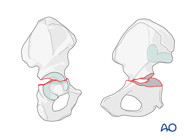
The posterior portion of transtectal transverse fractures often extends cranial to the superior gluteal neurovascular bundle. Exposure to this region of the fracture may be limited through the traditional Kocher-Langenbeck approach.
Prof. Letournel described the transtectal transverse fracture as an indication for the use of the extended iliofemoral exposure for this reason. Depending on the exact relationship of the fracture to the radiological roof, extension of the Kocher-Langenbeck with a trochanteric osteotomy or use of a true surgical dislocation may allow the surgeon sufficient access to reduce many transtectal transverse fractures, rather than resorting to the extended iliofemoral approach.
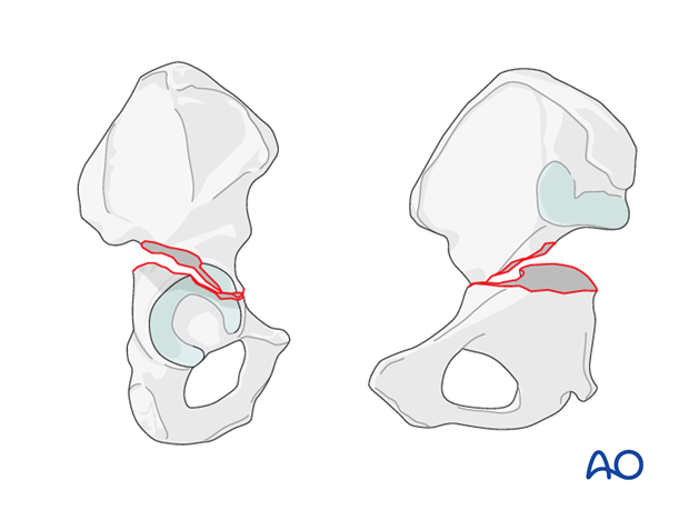
In rare cases, the anterior column of the ischiopubic segment is more displaced than the posterior. In these rare cases, an ilioinguinal or modified Stoppa exposure may be used to directly reduce the anterior ischiopubic segment. The posterior column reduction through the anterior exposure is attempted with various clamp applications, and fixation with anterior to posterior column screws or quadrilateral plate implants may be possible. Alternatively, if the posterior column is insufficiently reduced, a subsequent Kocher-Langenbeck may be required.
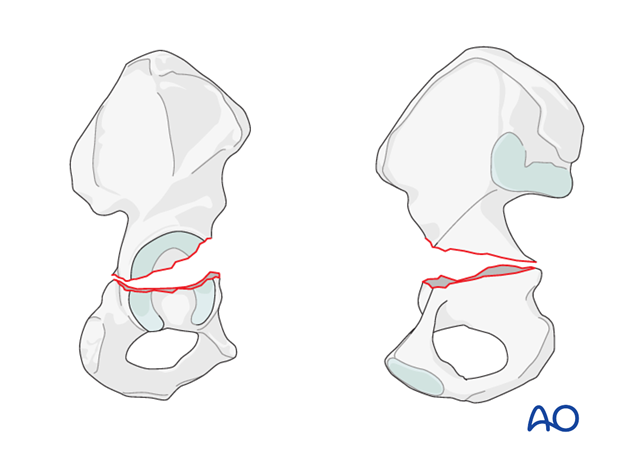
3. Kocher-Langenbeck approach
Indications
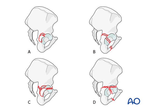
- Most posterior wall fractures (A)
- Posterior column fractures (B)
- Most transverse fractures with or without posterior wall fracture (C)
- Associated fractures in which the posterior column or wall requires reduction under direct visualization (certain associated both column and T-shaped fractures) (D)
Exposure
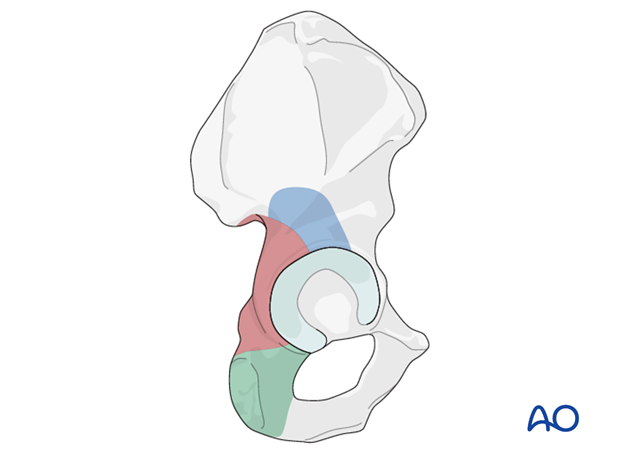
The Kocher-Langenbeck approach, in its native form, allows exposure of the retroacetabular surface as illustrated in dark brown. This is sufficient access for reduction and stabilization of most posterior wall fragments.
Additional exposure to the cranial anterior portion of the acetabulum (blue) can be obtained with trochanteric osteotomy.
Caudal exposure (green) can be obtained with reflection of the quadratus femoris muscle.
4. Trochanter osteotomy extension
Indications
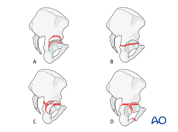
The most common use for this approach variant is the posterosuperior wall fracture (A).
In addition, certain transverse (B), transverse plus posterior wall (C), T-shaped (D), and associated posterior column plus posterior wall fracture patterns may be treated using this approach.
Factors which would favor this alternative include superior wall extension or a transtectal transverse fracture component in a patient thought to be a poor candidate for an extensile or combined approach.
Exposure
The addition of the trochanter osteotomy to the Kocher-Langenbeck or Gibson exposure provides improved access to the cranial and anterior aspects of the acetabulum and supra-acetabular surface.
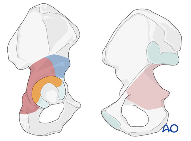
The dark brown area demonstrates the exposure associated with the osteotomy and the patient positioned prone.
The blue region demonstrated the additional exposure possible through the Gibson interval with the patient in a lateral position. The more anterior interval and the additional hip motion allow exposure to the anterior inferior iliac spine, not possible with the patient prone.
The intraarticular region outlined in orange demonstrates the additional exposure associated with complete hip dislocation.
The area in light brown can be accessed by palpation and allow clamp placement.
5. Anterior approaches
Indications
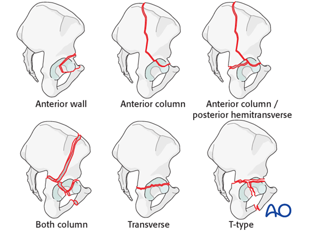
The modified Stoppa approach or the ilioinguinal approach are used for virtually all fractures of the anterior column as well as associated anterior plus posterior hemitransverse patterns.
The majority of both column fractures, an occasional transverse or T-shape fracture can be treated using these approaches.
Modified Stoppa vs ilioinguinal approach
The modified Stoppa approach allows better control of the periacetabular column than the ilioinguinal approach, such that instead of beginning the reduction at the crest and then using the pelvic brim plate to neutralize the reduction, the pelvic brim plate is used to drive the reduction as well as stabilize it.
The Stoppa window does not allow easy visualization and instrumentation of the anterior wall.
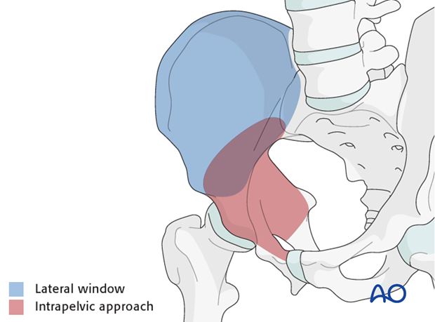
Exposure of the modified Stoppa approach
This approach to the anterior intrapelvic region (brown) allows visualization of:
- The pubic symphysis
- The entire quadrilateral surface
- The entire anterior column when combined with a lateral window (blue)
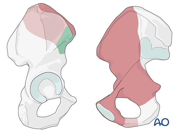
Exposure of the ilioinguinal approach
The approach allows direct access to the vast majority in the anterior ilium as depicted in dark brown.
Extended access of the outer ilium can be achieved by elevating limited portions of the tensor fascia latae origin (green) as well as the glutea (light brown). Additionally, clamps may be applied submuscularly in these regions to limit additional muscular trauma.
6. Extended iliofemoral approach
Indications
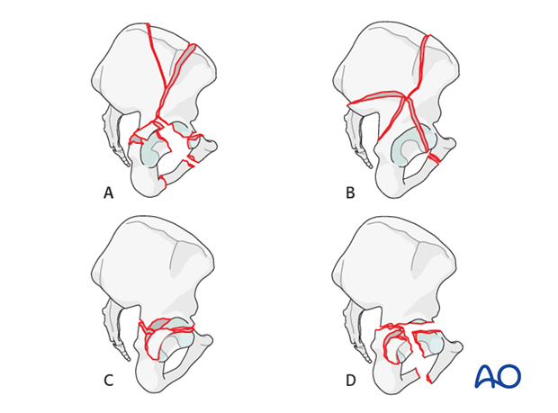
- Both column fractures with posterior wall or posterior column comminution, SI joint involvement (A), or very high posterior column involvement (B)
- Some transtectal associated transverse + posterior wall fractures (C), or T-shaped fractures, particularly with posterior wall comminution (D)
- Some transtectal T-shaped fractures with widely displaced vertical limbs or pubic symphysis dislocation (D)
- When ORIF of associated or transverse fractures is delayed by three or more weeks
Contraindications
The extended iliofemoral approach should not be used in aged or obese patients, or in patients who are not committed to a long recovery process.
The extended iliofemoral approach may be contra-indicated in patients who have undergone coil embolization of the ipsilateral superior gluteal arterial tree.
Advantages
The extended iliofemoral approach allows simultaneous visualization of both posterior and anterior columns, as well as intra-articular access. With this exposure, reduction and fixation are relatively straightforward due to the vast exposure of the innominate bone.
Disadvantages
Technically, this approach is demanding and has the highest complication rate. Heterotopic bone formation is common (prophylaxis should be planned). Abductor muscle weakness with prolonged rehabilitation must be expected.
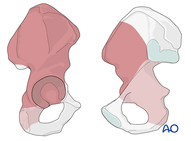
Exposure
This approach allows direct access to the area indicated in dark brown. The area in light brown can be accessed by palpation and allows clamp placement.
Extended iliofemoral approach vs sequential approaches
The extended iliofemoral approach has the highest complication rate (infection, heterotopic bone formation, neurological injury), and is technically the most demanding of the surgical approaches.
Its use is therefore reserved for fractures which, due to morphological complexities, cannot be reduced via an anterior exposure alone.
For some surgeons, the risk of wound breakdown is sufficiently great that they would choose two approaches over this single, combined approach. A combination of the modified Stoppa approach and the Kocher-Langenbeck approach in the prone position is the easiest and most reliable combination.
Both column or T-shaped fractures would be the most common fractures to treat with a sequential approach.
Case examples for extended iliofemoral approach
Complicating factors of associated both column fractures that reduces the chance of an adequate reduction through an anterior approach alone include:
- Complex posterior column involvement (A)
- Associated SI joint fracture dislocation (B)
- Large, displaced, or atypical posterior wall fragments (C)
- Wide displacement at the acetabular rim (not shown)
These morphologies may require direct access to the external surfaces of the bone and the posterior column for sufficient reduction and fixation.
