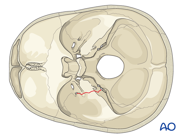
Skull base fracture, temporal bone
Introduction
Throughout human evolution, traumas severe enough to fracture the temporal bone were probably not compatible with survival. However, with the advances of modern medicine people are more likely to survive these injuries. Therefore, familiarity with their diagnosis and management is important.
Anatomy
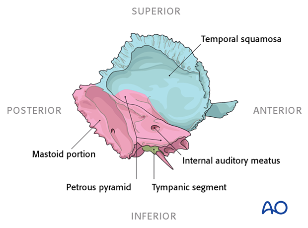
- Temporal squamosa
- Tympanic segment (ring housing ear canal)
- Mastoid
- Petrous portion (pyramid)
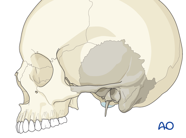
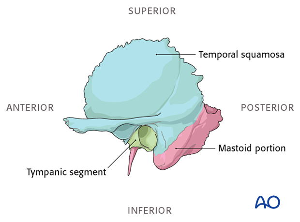
From the lateral aspect the largest portion seen is the temporal squamosa (squamous part) which occupies the superior two thirds of the illustration. As its name implies, it is the plate like portion of the bone that forms part of the cranium. Anteriorly the zygomatic root is formed by an anterior extension that juts out. The mastoid sits just posterior to the tympanic ring which forms the bony portion of the external auditory canal. The styloid process is seen projecting inferiorly just anterior and medial to the tympanic ring. Since the petrous portion projects medially, it is barely seen from this view.
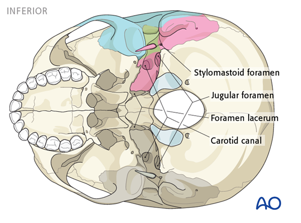
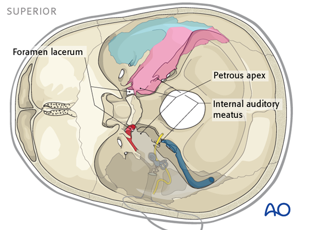
From inferiorly, you see the important openings in the skull base, such as the foramen lacerum and carotid canal (internal carotid artery), the jugular foramen (internal jugular vein, CN IX, X, XI), and the stylomastoid foramen (CN VII).
The medial view demonstrates the temporal bone from the intracranial perspective. Note that the somewhat anterior angulation of the petrous apex makes the internal auditory meatus (porus acousticus) visible from this view.
From the superior view you see that the temporal bones form the floor of the middle cranial fossa.
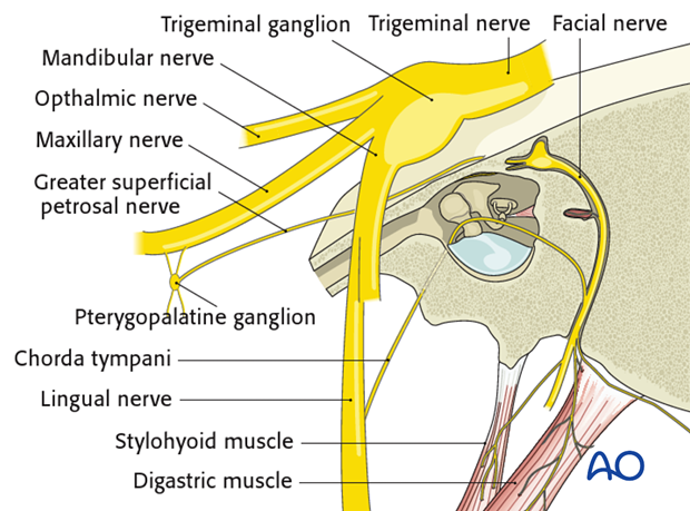
The relationship between the facial nerve in the temporal bone and the trigeminal nerve running across the petrous apex is shown. It depicts how the chorda tympani comes off the vertical portion of the facial nerve, to join the third division of the trigeminal nerve after exiting the middle ear through the anterior iter.
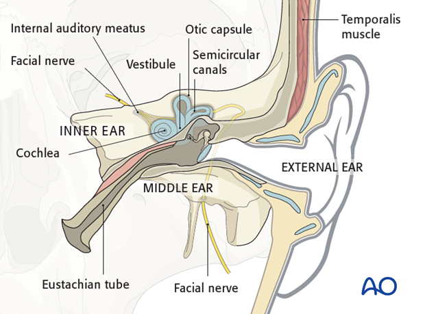
The important structures of the external ear, the middle ear and the inner ear are shown, depicting their relationship to the temporal bone.
This demonstrates the many vital structures that can be injured when the temporal bone is fractured. The cochlea, vestibule, and semicircular canals are housed in the temporal bone within the otic capsule.
Mechanism of the injury
Most temporal bone fractures are the result of large impact forces and therefore as might be expected they are most frequently the result of motor vehicle accidents and other high velocity impacts.
Clinical presentation
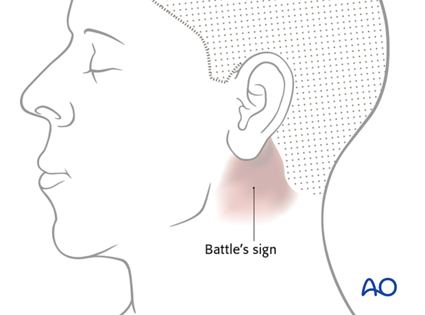
The presence of bloody discharge from the ear or ecchymosis behind the ear is suggestive of a temporal bone fracture.
Patients who are awake may complain of hearing loss, vertigo, facial weakness/paralysis, tinnitus, and/or taste dysfunction.
When there is blood coming from the ear canal, it is important to recognize if it is thinner than typical blood and might represent CSF otorrhea. It should also be noted that a CSF leak into the middle ear may present as rhinorrhea (otorhinorrhea) since it may pass through the Eustachian tube and drain out the nose. CSF leakage must be identified since it poses high risk for meningitis.
It is important to identify the presence of a facial nerve injury as soon as possible since most surgeons believe that delayed onset of a paralysis provides an excellent prognosis for recovery of the nerve and therefore contraindicates surgical decompression. Immediate paralysis on the other hand is an indication for further diagnostic work up as this may be an indication for surgery in selected cases.
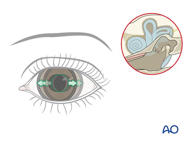
The presence of nystagmus is suggestive of vestibular trauma. Examination may reveal a perforated tympanic membrane and/or laceration of the ear canal. If the ear drum is intact, blood may be seen behind it (hemotympanum).
Imaging
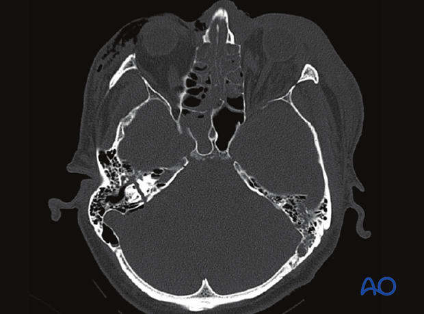
This CT scan shows a longitudinal temporal bone fracture.
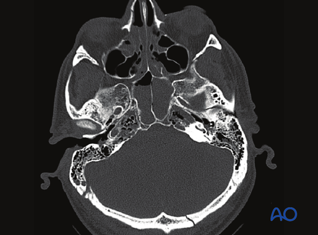
CT scan showing a transverse temporal bone fracture.
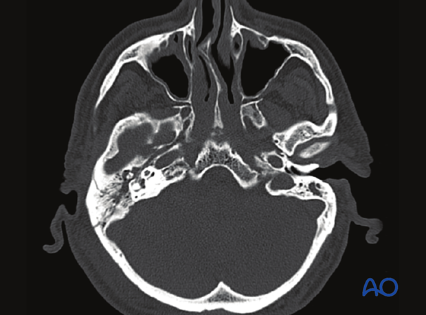
CT scan showing another transverse temporal bone fracture.
Classification
The classic description of temporal bone fractures comprises longitudinal, transverse and mixed fractures depending upon the relationship of the fracture to the long axis of the petrous apex. Clinical outcomes suggest that transverse fractures are associated with higher impact injuries and predict a worse clinical outcome. They are also associated with a higher incidence of facial nerve and otic capsule injuries. More modern classification systems have been proposed that are more clinically relevant, such as whether or not the otic capsule is involved or based on the lateral to medial extent. Involvement of the otic capsule is an excellent predictor of facial paralysis, CSF leak, hearing loss, and other intracranial complications.
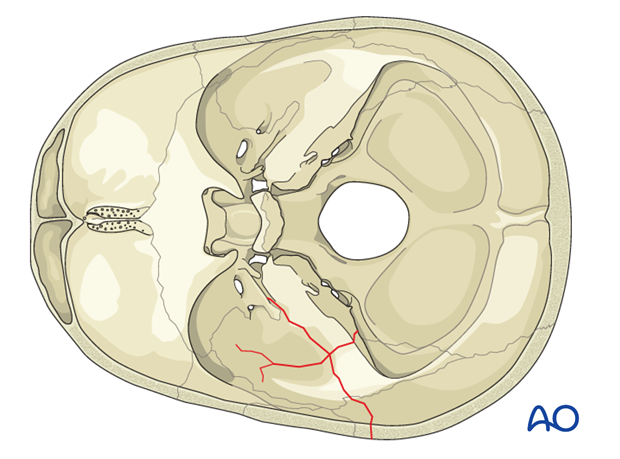
Illustration shows a longitudinal temporal bone fracture.
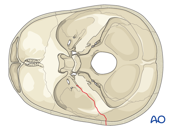
Illustration shows a transverse temporal bone fracture.
