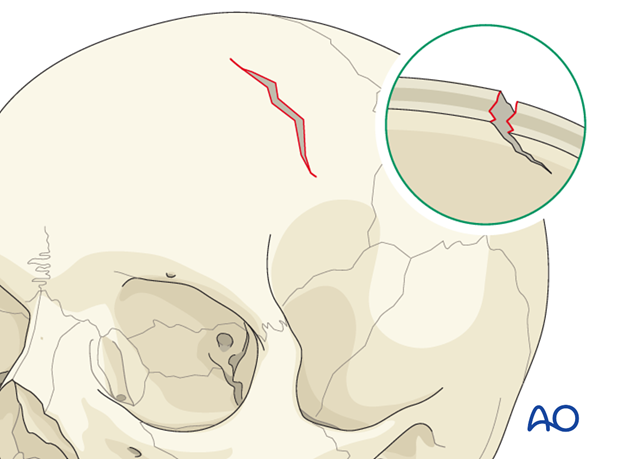
Skull base fracture, anterior
Introduction
Skull base fractures are of high importance in neurotrauma. They occur in 3.5 - 24% of head injuries and are often related to brain injury (in 50% of the cases).
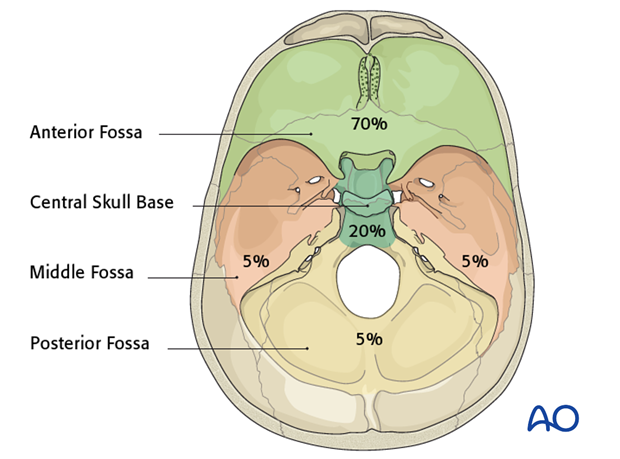
70% of the skull base fractures occur in the anterior fossa, 20% in the middle central skull base and 5% in the middle and posterior fossa.
Traumatic (CSF) leakage
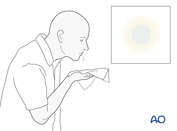
The most relevant clinical sign related to skull base fractures is CSF leakage. It occurs in 2% of all head trauma and can reach 30% of all skull base fracture cases.
80% of the traumatic CSF leakage occurs within 48 hours after injury.
16% of cases are “occult“, being found after recurrent meningitis.
Anatomy
Introduction
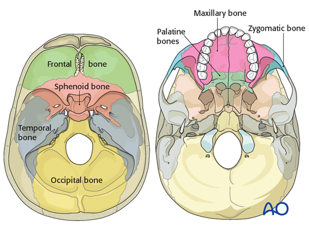
We consider the endocranial (inner) surface of the skull base, which consists of the cranial cavity on which the brain rests, and the exocranial (external) surface. The bones which form the skull base are:
- Frontal bone
- Sphenoid bone
- Temporal bone
- Occipital bone
The anterior part of the exocranial surface is also formed by the:
- Zygomatic bone
- Maxillary bone
- Palatine bones
The bones of the skull base contain several foramina through which nerves, arteries, and veins pass.
Anatomically, the inner surface of the skull base is formed by:
- Anterior fossa
- Middle fossa
- Posterior fossa
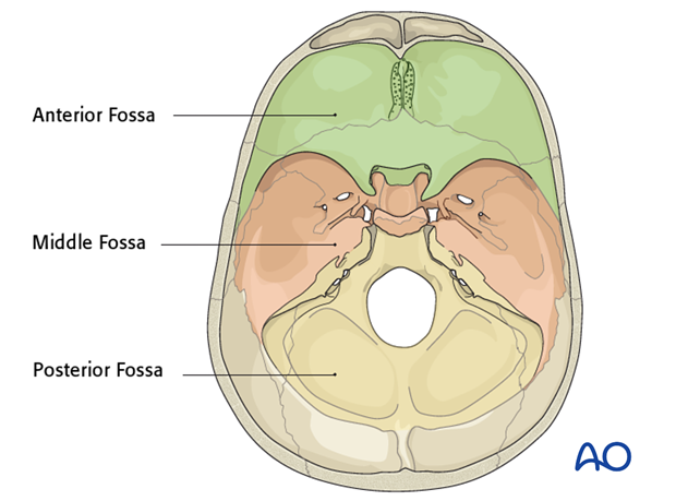
Anterior fossa
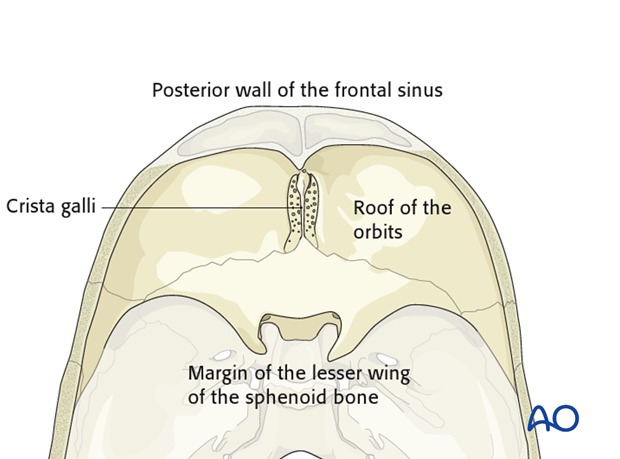
The anterior fossa is formed by the ethmoid bone, sphenoid bone and frontal bone. It is limited anteriorly by the frontal bone and the posterior wall of the frontal sinus, posteriorly by the limen of the lesser wing of the sphenoid bone. The lateral parts form the roof of the orbits. The median (central) part is formed by the crista galli, the cribriform plate of the ethmoid plane and the planum of the sphenoid bone.
Middle fossa
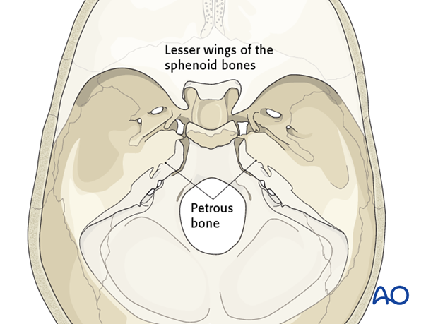
The middle fossa is formed by the sphenoid and temporal bones. It is limited anteriorly by the lesser wings of the sphenoid bones, posteriorly by the petrous bones.
Posterior fossa
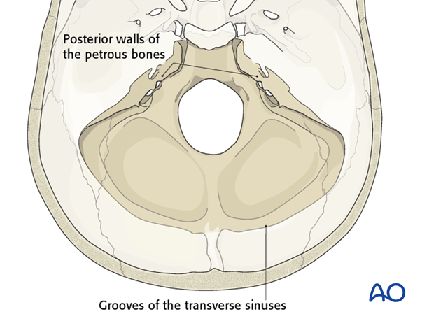
The posterior fossa is formed by the occipital bones. It is limited anteriorly by the posterior walls of the petrous bones and posteriorly by the grooves of the transverse sinuses.
Extension of anatomical classification
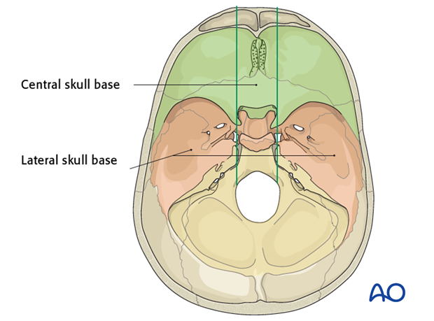
By drawing two horizontal lines which reach the lateral margins of the optic canals, the skull base can be divided into three longitudinal regions:
- Central skull base
- Lateral skull base (left and right)
Thereby, the inner surface of the skull base is divided into 9 quadrants.
Central skull base
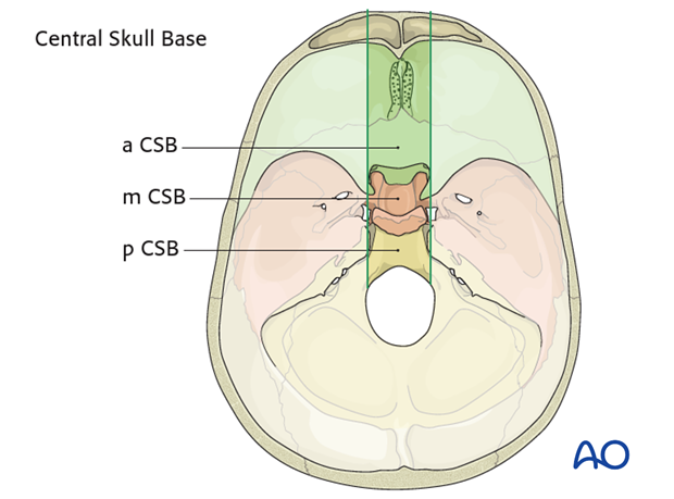
The anterior central skull base (a CSB) covers the upper nasal cavity and the sphenoid sinus.
The middle central skull base (m CSB) contains laterally the cavernous sinuses with the carotid arteries inside (parasellar compartments).
The posterior central skull base (p CSB) includes the clivus reaching the anterior margin of the great occipital foramen.
Cranial nerves and related skull base foramina
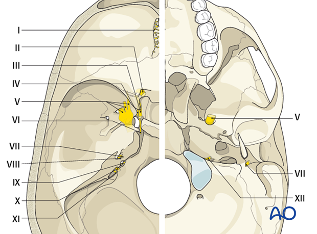
When fractures involve some specific anatomical regions the involvement of nerves passing through a foramen in the respective region should be always considered.
I Olfactory nerve: formed by many sensory nerve fibers that extend from the olfactory epithelium to the olfactory bulbs passing through the openings of the cribriform plates of the ethmoid bone (in the anterior central skull base).
II Optic nerve: passes from the retina to the brain in the optic canal in close relationship with the anterior clinoid process (middle central skull base).
III Oculomotor nerve: enters the orbit through the superior orbital fissure between the middle and anterior fossae.
IV Trochlear nerve: enters the orbit through the superior orbital fissure between the middle and anterior fossae.
V Trigeminal nerve: is made up of three divisions:
- Ophthalmic branch which passes through the superior orbital fissure
- Maxillary branch which passes through the foramen rotundum
- Mandibular branch which passes through the foramen ovale
VI Abducens nerve: enters the orbit through the superior orbital fissure between the middle and anterior fossae.
VII Facial nerve: enters the petrous temporal bone via the internal auditory meatus and emerges from the external surface of the skull base through the stylomastoid foramen (lateral posterior skull base)
VIII Vestibulocochlear nerve: enters the internal acoustic meatus.
IX Glossopharyngeal nerve: passes the through the jugular foramen.
X Vagus nerve: passes the through the jugular foramen.
XI Accessory nerve: starts outside the skull, enters the skull through the foramen magnum and exits again with the IX and X nerve through the jugular foramen.
XII Hypoglossal nerve: passes through the hypoglossal canal in the occipital bone.
Extracranial surface
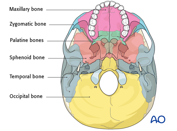
The extracranial surface is formed by:
- Occipital bones
- Temporal bones
- Sphenoid bones
- Palatine bones
- Zygomatic bones
The specific structures that can be involved in fractures of the extracranial surface of the skull base are the:
- Styloid processes of the temporal bone
- Tips of the mastoid bones
- Occipital condylar processes
Mechanism of the injury
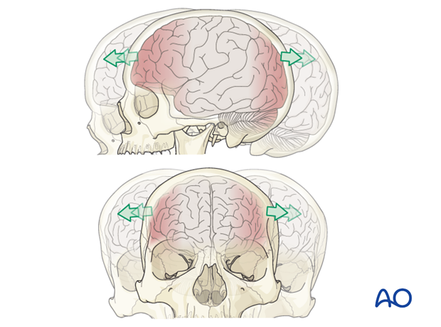
The skull base is particularly susceptible to the effects of blunt trauma. Skull base fractures are often associated with cranial vault or midface fractures.
The most vulnerable regions of the skull base are the petrous bone, the sphenoid sinus, and the foramen magnum.
Clinical presentation
Since skull base fractures are the results of high force impacts and are often associated with other intracranial injuries. Therefore, patients may be unconscious or require intervention for other more life-threatening injuries. As a result, the clinical signs and symptoms of skull base fractures may not be recognized immediately.
Patients affected by skull base fractures can present anywhere from awake and asymptomatic to comatose or even moribund.
The first clinical assessment is the evaluation of the Glasgow coma scale (GCS).
It is important to recognize the blood and/or CSF coming from the ear (otorrhea), the nose (rhinorrhea), or some calvarial wounds. CSF leakage must be identified since it poses high risk for meningitis. For suspected but not evident rhinorrhea a provocation test (Valsalva maneuver) can be useful. Other useful test can be:
- Double ring sign
- Glucose test strip
- Beta-2-transferrin test

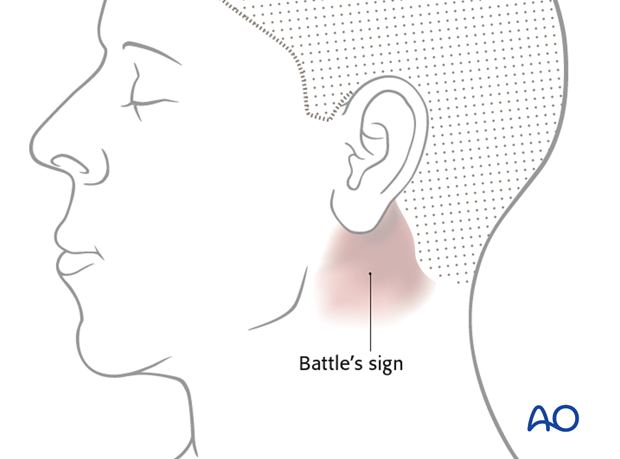
The presence of subcutaneous ecchymosis in the mastoid region (Battles’ sign) or ...
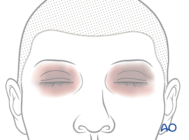
... around the eyes (raccoon’s eyes) is very highly suspicious for skull base fractures.
In awake patients it is important to identify the presence of cranial nerve injury as soon as possible especially of the optic and facial nerves.
A complete neurological examination has to be done in all cases.
Imaging
The gold standard for the radiographic detection of skull base fractures is computed tomography.
Specific, very useful CT sequences are:
- Non contrast high resolution bone window CCT (thin slices 1mm, axial and coronal)
- Multiplanar reconstructions
Special modalities include:
- MRI
- Cerebral angiography
- CT-cisternography
Classification
Single (linear and/or branched) and multiple
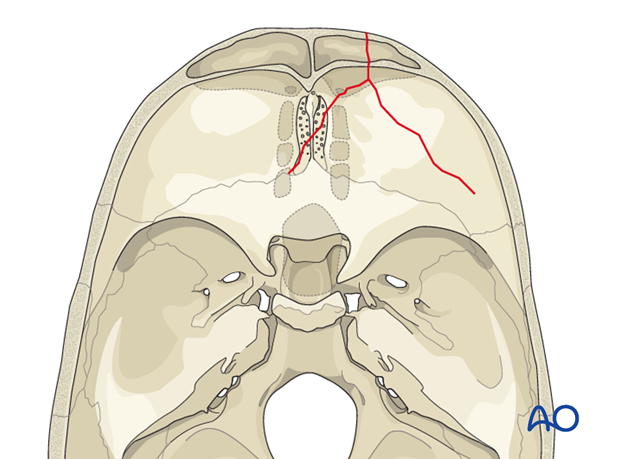
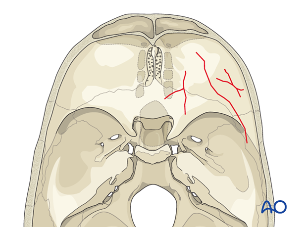
The fractures can be single, crossing more bones, or multiple, in the same bone or in different bones. The fracture can be linear or branched.
Single fracture line
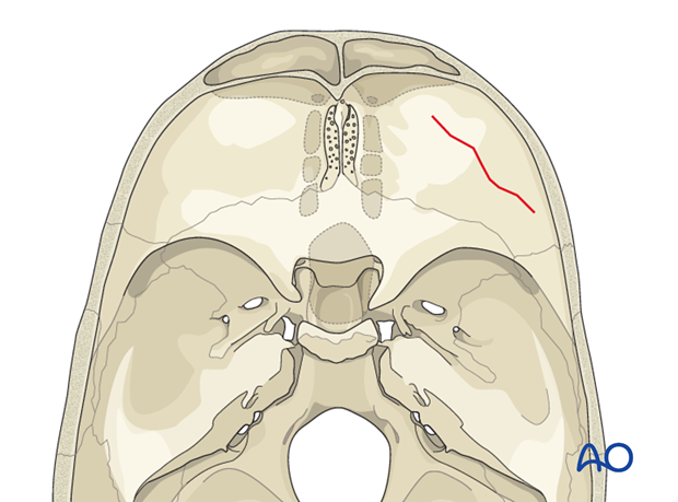
Branched fracture lines.
Multiple separated fracture lines.
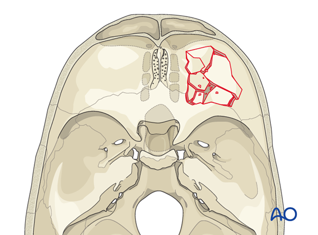
Comminuted
A fracture is comminuted when the bone is shattered into many fragments.
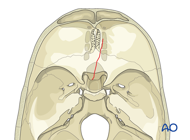
Contiguous
The fracture is contiguous when it crosses anatomical boundaries.
Depressed
The fractured segments are displaced inward, toward the meninges and brain for more than 3 mm.
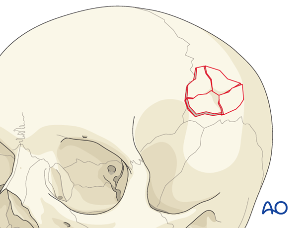
Diastatic suture
Horizontal displacement along the cranial sutures (>3 mm).
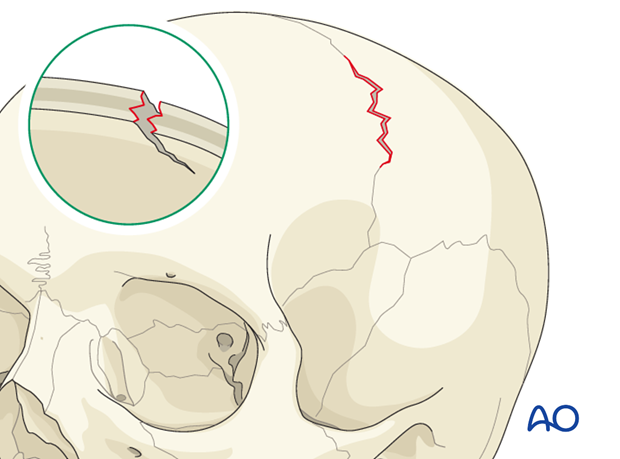
Diastatic fracture
Horizontal displacement of the bones at the margin of the fracture (>3 mm).
