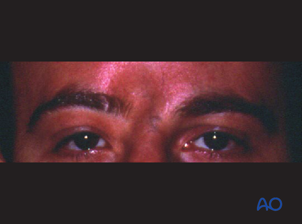
Complications
1. CNS Infections
Meningitis/Encephalitis
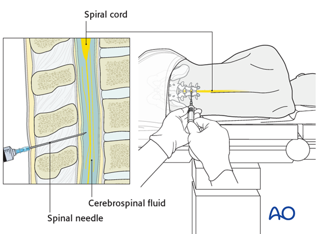
Meningitis is due to ascending infection in the presence of a dural defect/CSF leak. Typical symptoms include fever, headache, stiffness of the neck, positive Lasègue's, Brudzinski, and Kernig sign, clouding of consciousness. Diagnosis is confirmed by CSF examination (lumbar puncture). Patients with meningitis require intensive care.
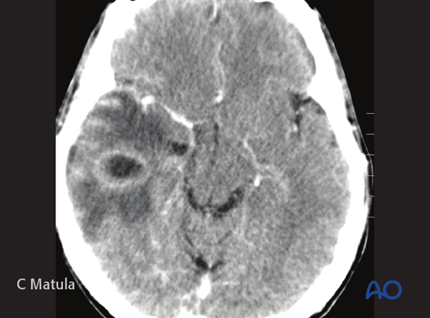
Brain abscess
Accumulation of pus intracranially requiring surgical intervention.
2. Paranasal sinus infections
Fronto-ethmoidal sinusitis
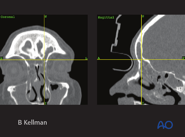
Fronto-ethmoidal sinusitis is due to obstructed drainage of the frontal and ethmoid sinuses. Initial treatment includes antibiotics and decongestions. Sometimes, surgical intervention may be indicated.
The availability of transnasal endoscopic sinus surgical techniques, particularly with the aid of image guidance (computerized navigation), allows for minimally invasive management of these problems.
3. Ophthalmological complications
Diplopia
Diplopia is a rare complication of cranial and skull base fractures.
Loss of vision
Visual loss may occur in case of fractures affecting the optic canal leading to traumatic optic neuropathy. Initial treatment is done with mega-dose steroids followed by surgical decompression even though both treatments are still controversial due to lack of scientific evidence.
Enophthalmos
Enophthalmos is not a typical complication of skull base/cranial vault fractures.
Exophthalmos, Dystopia
This may be caused by dislocated orbital roof fractures. Treatment may require a coronal approach and an orbital osteotomy and/or craniotomy.
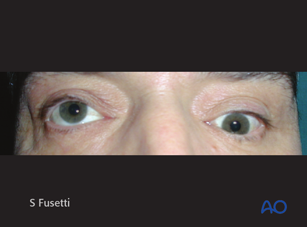
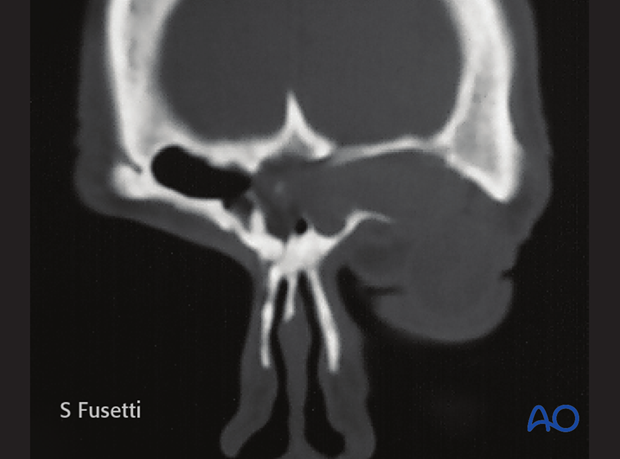
Late exophthalmos and/or hypophthalmos may result from an expanding frontal mucocele/mucopyocele that has invaded the orbit.
Eyelid malposition
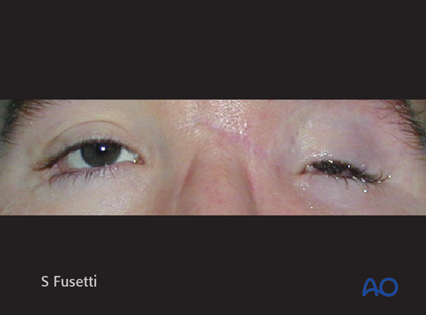
Eyelid malposition due to incorrect positioning of the medial canthal tendon is a frequent complication of displaced NOE fractures.
It is not a typical complication of skull base/cranial vault fractures. Ptosis could also be a result of neurologic injury.
Carotid-cavernous fistula
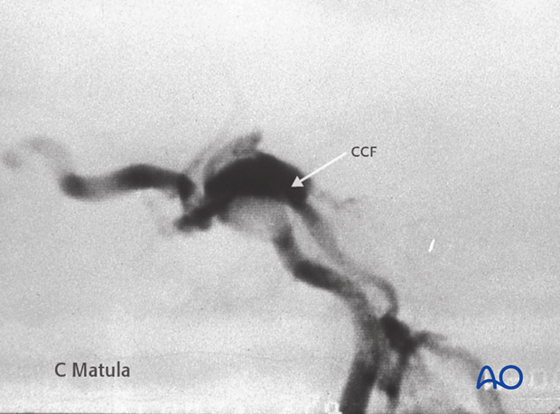
Carotid-cavernous fistula can cause ophthalmological complications and may result in pulsatile exophthalmos.
This complication may require endovascular treatment.
4. Anosmia
If the cribriform plate is fractured, patients may develop anosmia due to injuries to the olfactory nerves. Fractures of the cribriform plate can be associated with anterior skull base, NOE, and frontal sinus fractures.
Anosmia can also be a complication of the surgical repair of the fractures.
5. Facial nerve dysfunction
Fractures of the temporal bone can result in facial nerve injury.
The severity of facial nerve dysfunction should be categorized according to the House-Brackmann scale. Further details regarding the treatment of facial nerve paralysis are available in the dedicated section of the AO Surgery Reference.
6. Hearing loss
Fractures of the temporal bone can result in hearing loss.
Hearing loss may result from damage to the cochlear and/or VIII nerve (neurosensory hearing loss), and it may also result from damage to the middle ear structures (conductive hearing loss). Blood behind the ear drum (hemotympanum) can also be a cause of temporary conductive hearing loss.
Hearing loss should be evaluated 6 weeks or more after the injury to allow time for hemotympanum to resolve (although it should be assessed sooner if surgical intervention is contemplated prior to that time).
7. Persistent CSF leak
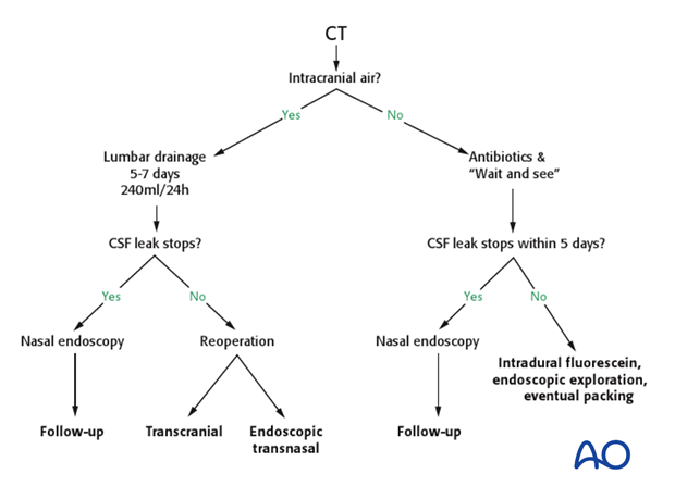
Recurrent and/or persistent CSF leaks can be difficult to repair.
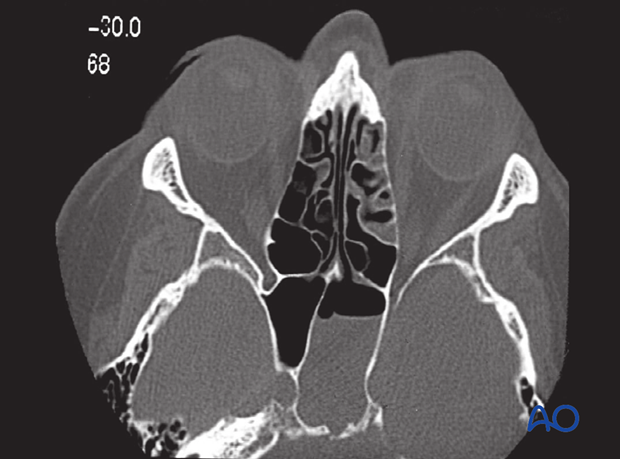
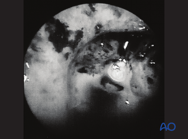
High definition CT scan examination and nasal endoscopy with intrathecal fluorescein injection should be used to determine the size and the site of the defect, respectively, thereby guiding the surgeon to the best approach to treatment. Transnasal repair is successful in a high percentage of cases and transcranial repair should be considered for very large defects or leaks that persist after endoscopic surgery has failed repeatedly.
CT scan by courtesy of Wolfgang Gstöttner, ENT Surgery, Vienna, Austria.
Nasal endoscopy with intrathecal fluorescein injection to help identify the site of the CSF leak.
Endoscopic photography by courtesy of Wolfgang Gstöttner, ENT Surgery, Vienna, Austria.
Treatment algorithm
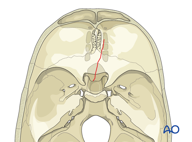
8. Complications of fractures involving the sellar region
Fractures extending to the sphenoid sinus/sellar region may cause dysfunction of the pituitary gland with subsequent diabetes insipidus.
In such cases, medical treatment is required.
9. Fractures of the middle fossa
Fractures of the middle fossa can cause oculomotor dysfunction. Fractures involving the superior orbital fissure or the foramina of the middle fossa can cause sensory trigeminal deficits.
In the latter condition, no specific treatments are available.
10. Occipitocervical instability
Fracture of the occipital condylar processes can affect the stability of the craniocervical junction.
In such cases, craniocervical fixation may be required.
11. Cosmetic deformities
Cranial vault fractures, if not properly treated by aligning the bony fragments or by reconstructing missing parts of bone can result in cosmetic contour deformities.
This photograph shows a right frontoparietal defect.
Photograph by courtesy of Umberto Zanetti, Brescia, Italy.
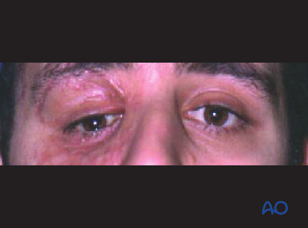
This photograph shows a depressed anterior table of the right frontal sinus and supraorbital rim.
Photograph by courtesy of Umberto Zanetti, Brescia, Italy.
