Cranial vault fracture
Introduction
The majority of head trauma involve the cranial vault.
Fractures most often occur in the frontal or parietal bone. In case of frontal vault fractures, typically the frontal sinus is involved.
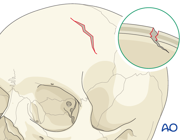
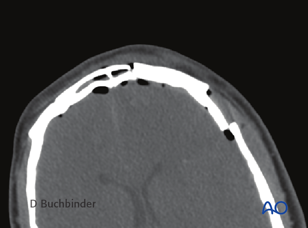
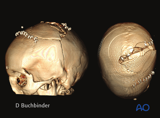
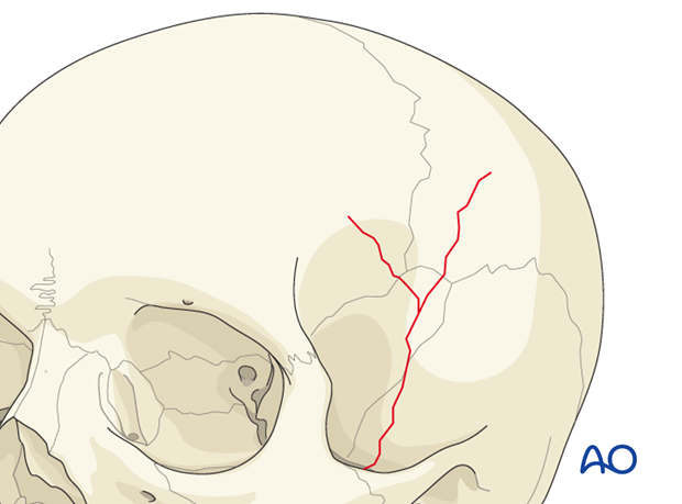
Many fractures can start between cranial sutures and extend to contiguous bones (eg, frontal, parietal, and temporal bones as illustrated).
The fractures of the cranial vault can be depressed, closed, or open with or without the involvement of the brain and meninges.
Anatomy
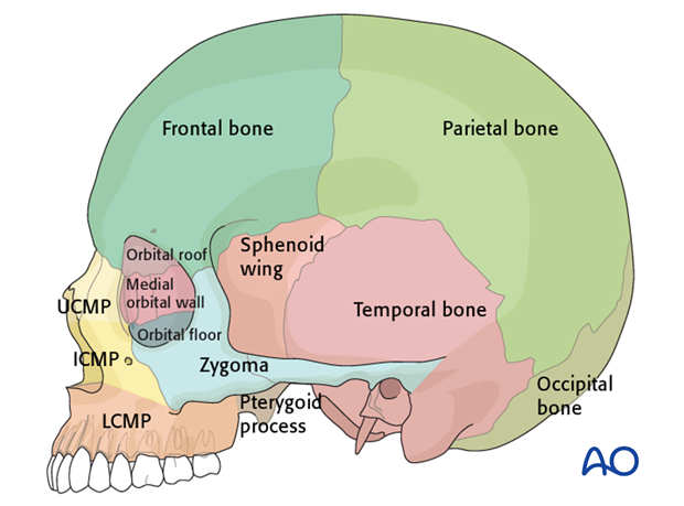
The cranial vault is formed by the frontal, parietal, occipital, and temporal bones, and the greater wings of the sphenoid bone.
Frontal Bone
A vertical portion, which corresponds to the forehead, and an horizontal portion which is part of the skull base, forming the roofs of the orbital and nasal cavities.
Parietal Bone
The two parietal bones form the sides and roof of the cranium.
Occipital Bone
The occipital bone forms the postero-lateral and posterior part of the cranial vault, more than the floor of the posterior skull base. It contains a central large oval opening, the foramen magnum.
Temporal Bone
The temporal bone has a petrous portion (part of the skull base), a lateral squama (forming the lateral cranial vault) and a mastoid portion.
Sphenoid Bone
Unpaired bone, of which the two lateral greater wings contribute to the formation of the anterior lateral cranial vault.
Mechanism of the injury
The most typical mechanism of cranial vault fractures are high velocity impact traumas. Cranial vault fractures are the result of a force that causes the skull to bend inward.
Clinical presentation and examination
The primary consideration in depressed closed or open skull fractures is the involvement of the brain and the meninges.
Patients affected by skull fractures can present awake, asymptomatic or comatose.
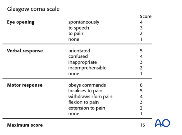
The first clinical assessment is the evaluation of the Glasgow coma scale (GCS).
The involvement of the meninges can lead to CSF leaks. If associated with frontal sinus fractures, typically a rhinorrhea is present. Cranial vault fractures can also cause entrance of air into the intracranial cavity (pneumocephalus). The latter condition rarely requires emergency treatment except in case of tension pneumocephalus in which a “ball valve” mechanism causes intracranial hypertension.
Fragments of cranial vault bones can cause brain damage (cortical lacerations) and/or vascular lesions (intra- or extracerebral hematomas, etc.). These conditions may require emergency surgery.
The clinician should examine for contracoup lesions (for example frontal trauma with a occipital contralateral lesion) as they frequently occur with skull fractures.
Imaging
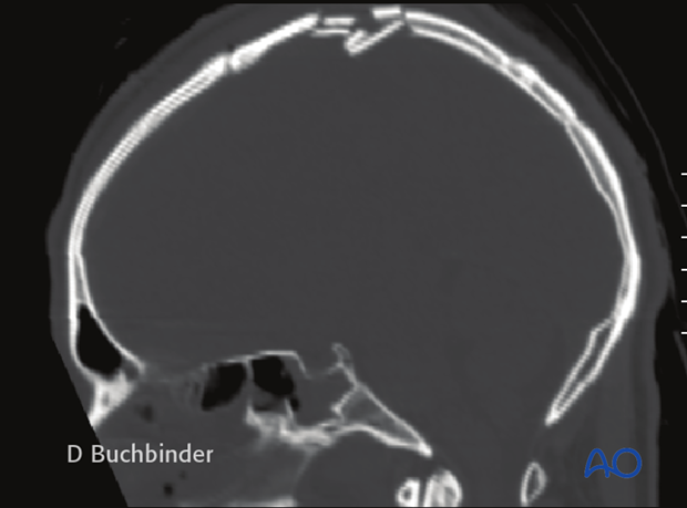
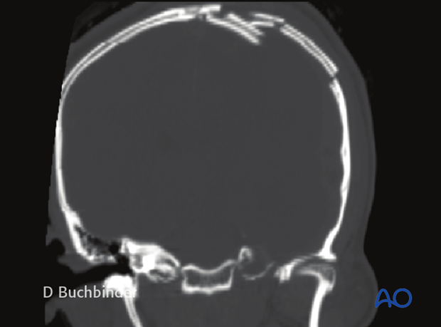
Although frontal bone fractures can be seen on plane x-rays, the gold standard for the radiographic detection of skull fractures is computer tomography (CT). A complete cranial and midface multiplanar CT-scan (should include cranial vault and skull base, orbits, sinuses, and temporal bones) should be obtained. Both bone and soft-tissue (brain) windows should be evaluated. Coronal sections are helpful in detecting fractures and assessing the degree of bone displacement.
Sagittal CT scan of a cranial vault fracture.
Coronal CT scan.
When available, 3-D reconstructions, and CT-Angiography (CTA) can be very useful.
Brain injuries (eg, lacerations, contusions, hematoma, etc), intracranial bleeding(traumatic subarachnoid hemorrhage, epi- sub-, or intracerebral hematoma) and intracranial air are also detected by CT imaging.
MRI examination is used for determining soft-tissue lesions which are not easily detectable by computed tomography.
Perfusion studies are rarely used. In selected cases, cerebral angiography can be helpful.
Classification
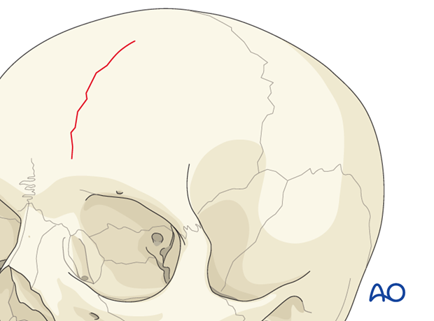
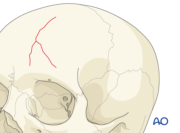
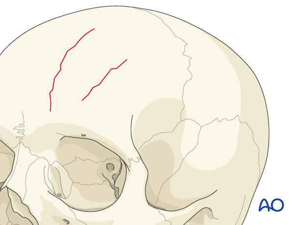
Single (linear and/or branched) and multiple
The fractures can be single, crossing to contiguous bones, or multiple, in the same bone or in contiguous bones. The fracture can be linear or branched.
Single fracture line.
Branched fracture lines.
Multiple fracture lines but not connected.
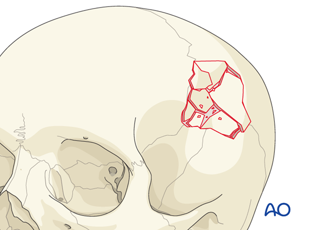
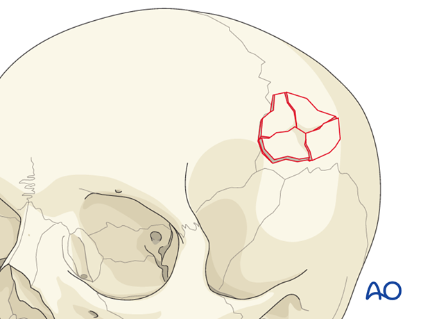
Comminuted
A fracture is comminuted when the bone is shattered into many fragments.
Contiguous
The fracture is contiguous when it crosses anatomical boundaries.
Depressed
The fractured segments are displaced inward, toward the meninges and brain for more than 3 mm.

Diastatic suture
Horizontal displacement along the cranial sutures (>3 mm).
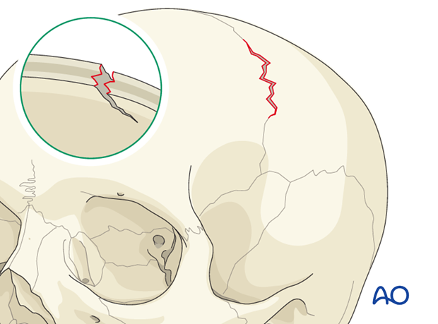
Diastatic fracture
Horizontal displacement of the bones at the margin of the fracture (>3 mm).
