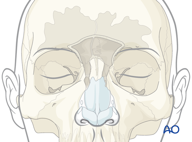
Endoscopic transnasal approach to the frontal sinus
1. Introduction
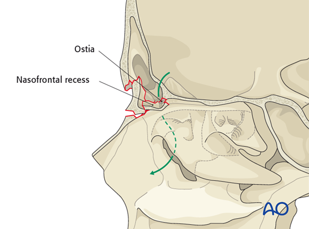
Injury to the frontal recess can result in obstruction of the outflow tract. While frontal sinus obliteration is a treatment option, endoscopic sinus surgery techniques can be used to open the frontal recess from below. This is a highly specialized technique reserved for surgeons with extensive experience in traditional endoscopic sinus surgery.
2. Frontal recess exposure
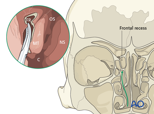
Resect the agar nasi cell and the anterior ethmoid air cells. The dissection is carried superiorly to circumferentially expose the frontal recess from below.
Legend:
FR: frontal recess
OS: ostium sphenoidalis
MT: middle turbinate
NS: nasal septum
C: Choana
3. Frontal sinus trephination
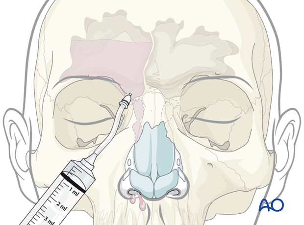
Frontal sinus trephination may be helpful for identification of the frontal sinus ostia. If the ostia is not completely blocked, irrigation of the sinus from above will reveal fluid drainage endoscopically from below. This can help define the location of the true ostia.
Intraoperative navigation can also be helpful in localizing the true ostia.
The surgeon must be aware that the skull base anatomy may be distorted secondary to the trauma.
4. Resection of septum
After isolation of the first sinus ostia, carry the dissection across the septum/midline. Use a scalpel and shaver to resect the superior 2-3 cm of the septum.

5. Enlargement of right frontal ostia
Enter the frontal ostia with a drill removing the nasal crest of the frontal bone. Move anterior and medially towards the contralateral ostia. Enlarge the true ostia to completely resect the nasal crest, expose the anterior skull base (posteriorly), and the medial orbital wall (laterally).
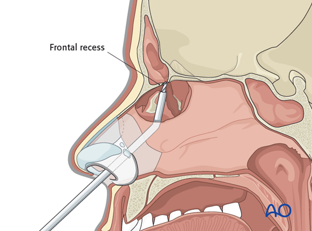
6. Enlargement of left frontal ostia
Use a similar technique to drill out the left frontal ostia.
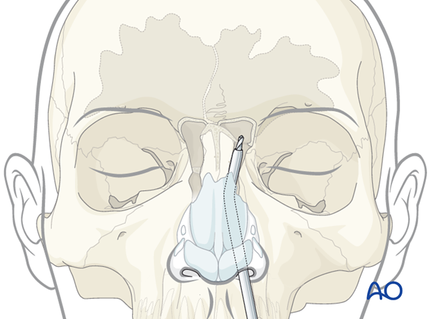
7. Resection of sinus floor
Drill across the midline to join the left and right sinus ostia. Remove as much bone as possible anteriorly and medially to enlarge the drainage pathway. Use caution when drilling posteriorly (skull base) and laterally (orbit).
