Endoscopic approach to the central skull base
1. Introduction
The whole central compartment of the skull base, from the crista galli to the clivus and anterior craniocervical junction, can be accessed by means of the endonasal transsphenoidal endoscopic approach.
2. Endonasal endoscopy
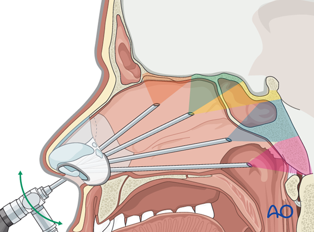
The endoscope is introduced in the nostrils and the main anatomical landmark should be identified. The 2 nostrils – 4 hands techniques is highly recommended: the collaboration of two surgeons (ENT and neurosurgeon) insure the best therapeutic option for the patients.
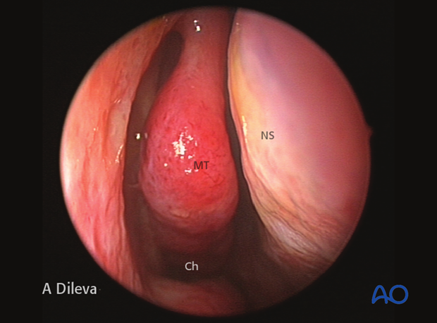
In the endonasal step, the main landmarks are: the middle turbinate (MT), the nasal septum (NS), the choana (Ch).
3. Finding of the ostium sphenoidalis
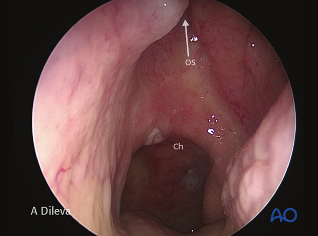
The roof of the choana (Ch) is a good anatomical landmark to find the ostium sphenoidalis (OS), which is located superiorly to it.
4. Opening of the ostium sphenoidalis
Once the ostium sphenoidalis (OS) has been identified, ...
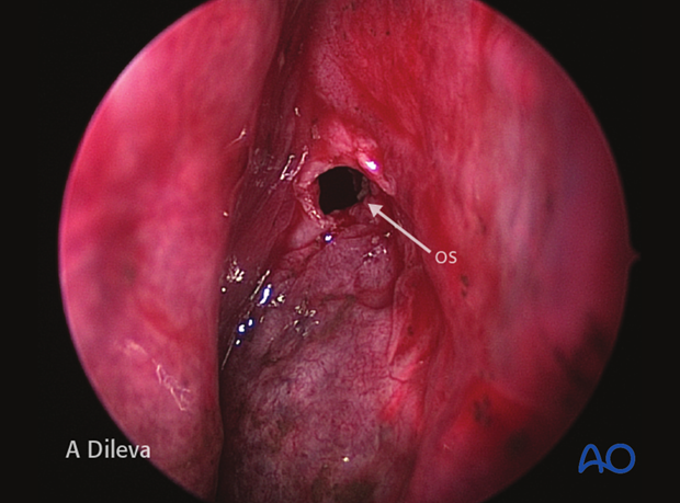
... it is enlarged using a diamond tipped burr and circular cutting punch.
According to the needs, the two openings of the right and left ostium sphenoidalis can be connected in order to get more space for surgical instrumentation.
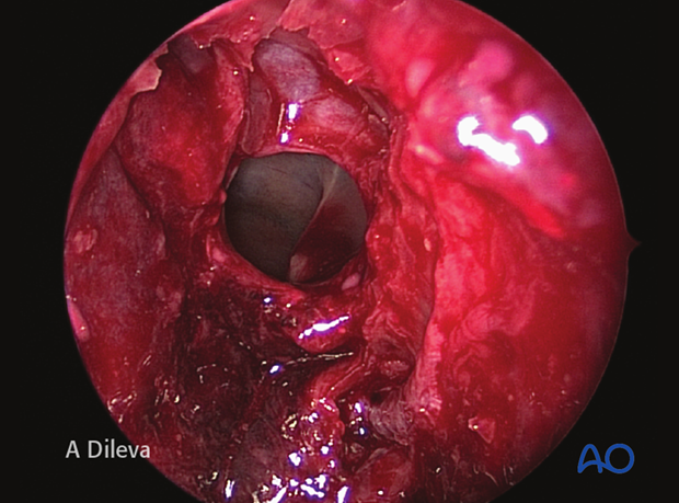
5. Sphenoidal step
The introduction of the endoscope in the enlarged ostium sphenoidalis allows the visualization of additional anatomical landmarks: sellar floor (SF), clivus (C), planum sphenoidalis (PS), internal carotid artery (ICA), and optic nerve (ON).
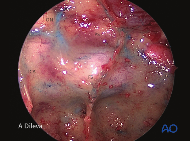
6. Visualization of the optic-carotid recess
The optic-carotid recess (OCR) is a very important landmark for the identification of the internal carotid artery (ICA) and the optic nerve (ON).
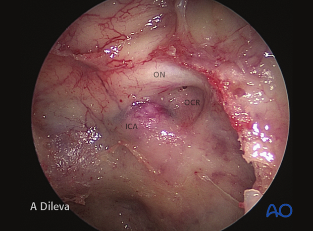
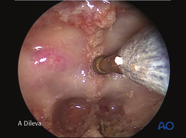
High-speed drills are used to open the floor of the sella (as in the photograph) and the planum sphenoidalis, to allow for decompression of the optic nerve and/or to allow for reduction of bony fragments, etc.
7. Extended endoscopic transbasal approach
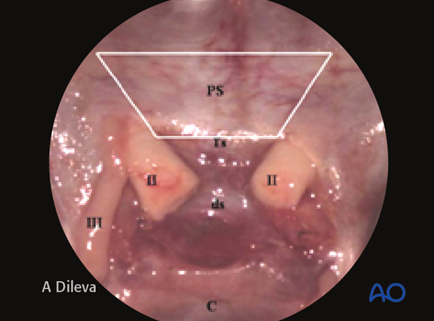
The anterior cranial fossa can be reached removing the planum sphenoidalis (PS) between the optic nerves (II), visualizing the dura mater (DM) of the basal frontal lobes. The endoscopic visualization shown here ...
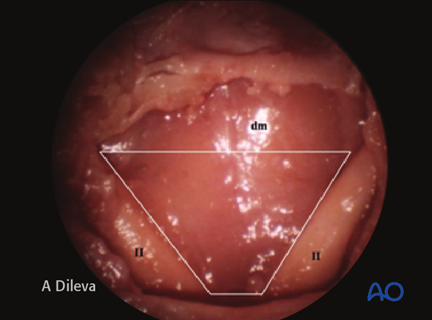
... is compared to the transcranial visualization of the same region. The area which has to be opened is outlined.
C: Clivus
III: oculomotor nerve
8. Duroplasty
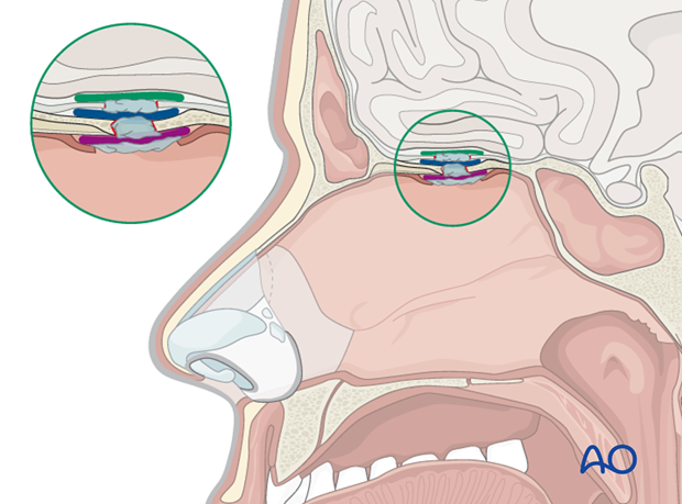
In cases of dural injuries, the closure technique is strictly related to the individual patient‘s anatomy, the size of the CSF leak, and its anatomical location. Underlay, overlay, combined, and obliterative techniques have been described.
The illustration shows a combined three layer technique in which are evident:
- Subdural intracranial underlay graft (dark green)
- Extradural intracranial underlay graft (blue)
- Extracranial overlay graft (purple)
Fibrin glue can be used to keep the layers together or to fill the dead-space.
