Endoscopic approach to the anterior table
1. Indications
Endoscopy has a variety of potential uses for frontal sinus fractures. It can be used for evaluation (trephination with sinus endoscopy), or for delayed endoscopic repair in patients with isolated fractures, limited to the anterior table who continue to have a cosmetic contour deformity after 3-4 months of observation and do not demonstrate any obstructive signs of sinus pathology. It can also be used to manage sinuses with obstructed outflow.
The reasoning and indications for delayed repair as described above should be articulated to the patient. It should be noted that a traditional reduction can not be performed secondarily.
An advantage of this approach is reduced patient morbidity (very limited incision with little risk of alopecia).
2. Access area
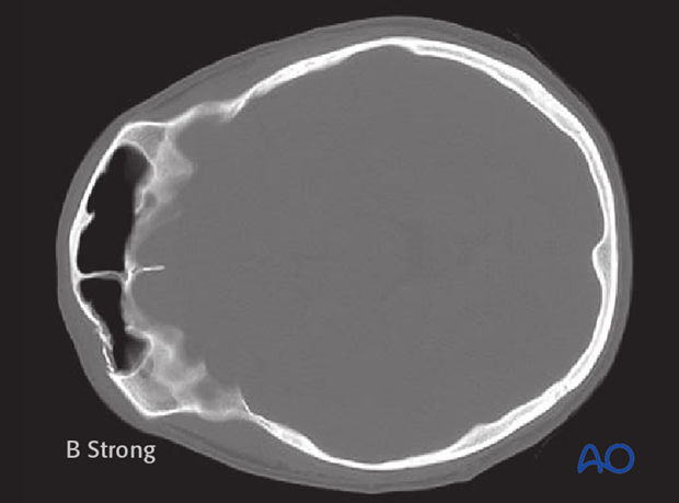
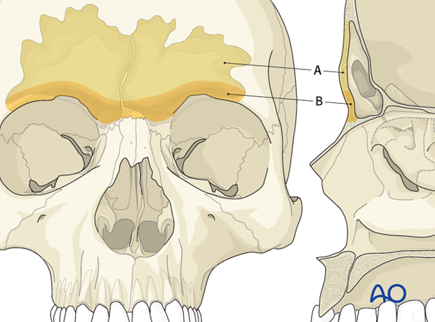
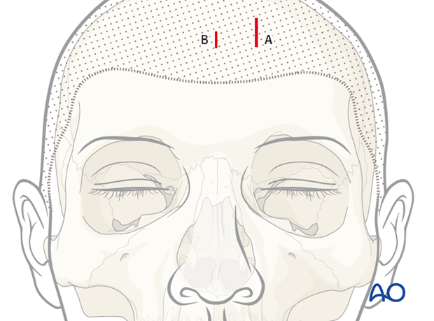
Endoscopic access is most favorable in the upper portion of the frontal sinus (A). Involvement of the orbital rims (B) is more difficult to manage endoscopically due to the intrinsic curvature of the skull.
3. Room set-up
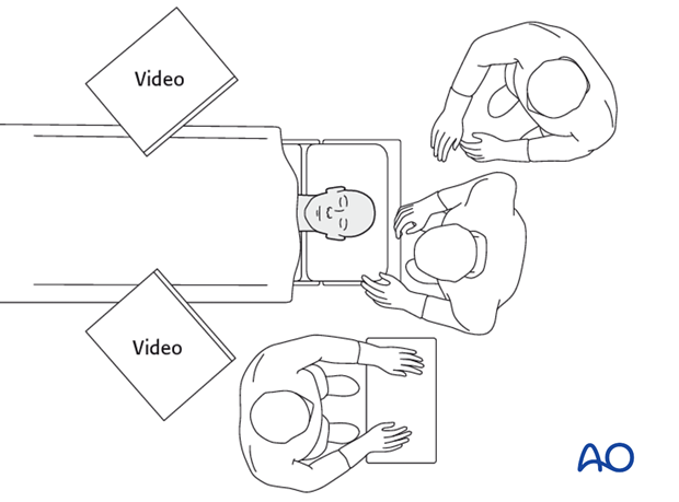
The OR table is turned 180°from the anesthesia cart. The endoscopic video cart(s) should be placed in such a way that they are readily visible to the surgeon and assistant surgeon.
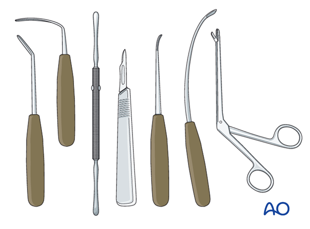
4. Instruments
A 30° 4 mm endoscope and endoscope sheath are used for visualization of the defect. A larger guard at the end of the sheath will maximize the optical cavity.
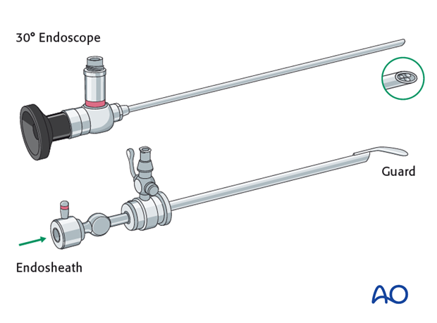
Typical browlift instrumentation is used for the endoscopic repair.
5. Skin incision
Local vasoconstrictors should be used liberally if possible and electrocautery should be avoided to reduce the risk of alopecia.
A 3 cm working incision is placed above the operative site at least 3 cm behind the hairline (A). This length will vary depending on the size of implant to be placed. An endoscope incision (B) is placed 4 cm medial to that, at least 3 cm behind the hairline. The incisions are carried down through the periosteum and onto bone.
Forehead incisions
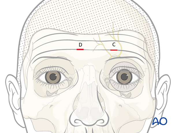
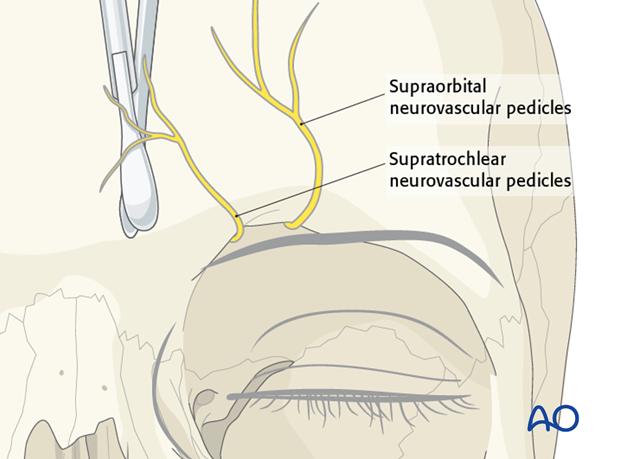
Skin incision should be aligned with natural skin creases, when possible. Caution should be used to avoid injury to the supratrochlear and supraorbital neurovascular pedicles.
Camouflage technique:
A stab incision is required directly over the defect for placement of implant fixation screw(s). (C)
Multiple stab incisions may be required when a larger implant is needed (C and D).
6. Tissue elevation
Browlift elevators are used to expose the defect in a subperiosteal plane. Blind dissection and bimanual palpation are used down to the level of the defect. Lacerations of the periosteum should be avoided because it will make insertion of the endoscope and visualization of the defect more difficult.
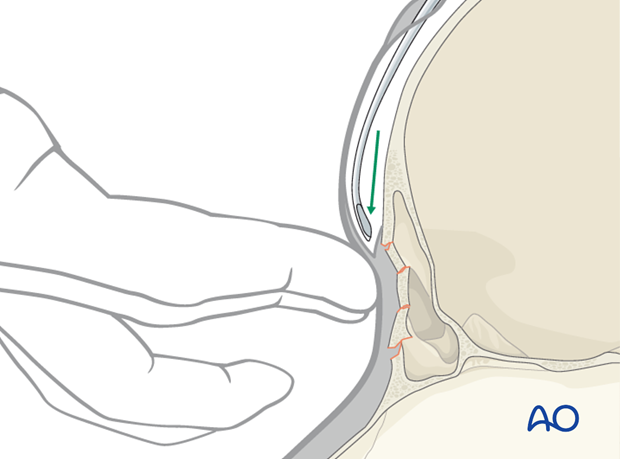
The endoscope is then inserted and the elevator is used to carefully expose the defect. The endoscope is generally held above the periosteal elevator by the assistant. Some experience is required to maintain endoscopic visualization without obstructing access for the periosteal elevator.
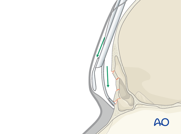
Caution should be used to avoid excessive traction on the supratrochlear and supraorbital neurovascular pedicles as this may result in postoperative paresthesia.
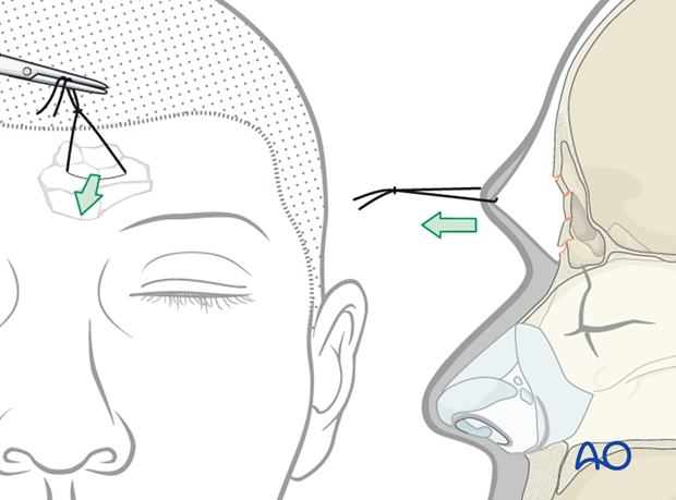
Pearl: increasing optical cavity size
A large (0 silk) suture can be passed full thickness through the forehead (skin, muscle, and periosteum) and traction placed to maximize the optical cavity.
7. Wound closure
The incisions are closed in layers and a pressure dressing is applied for 3 – 5 days.


