Zygoma, zygomatic complex fracture
Introduction
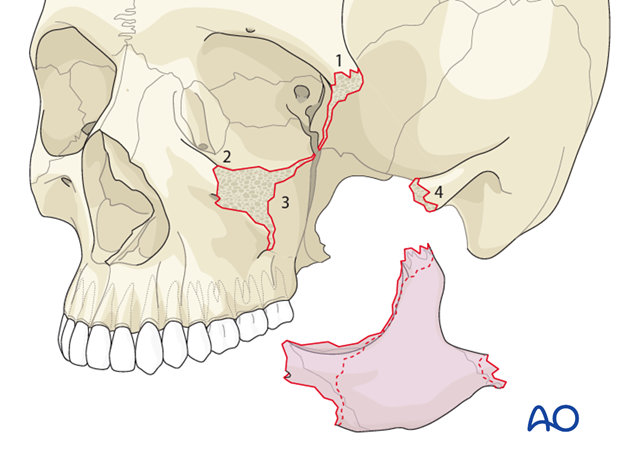
A zygomatic complex fracture is a fracture that involves the zygoma and its surrounding bones. The typical lines of a zygomatic complex fracture are:
- A fracture emanating from the inferior orbital fissure superiorly along the sphenozygomatic suture to the frontozygomatic suture where it crosses the lateral orbital rim
- A fracture emanating from the inferior orbital fissure anteriorly along the orbital plate of the maxilla, crossing the infraorbital rim and extending inferiorly along the anterior face of the maxilla underneath the zygomaticomaxillary buttress
- A fracture emanating from the inferior orbital fissure passing inferiorly along the infratemporal surface of the maxilla, passing anteriorly underneath the zygomaticomaxillary buttress to meet fracture 2 above
- One or more fractures through the zygomatic arch.
Radiographic findings
3-D reconstruction of a right zygomatic complex fracture.
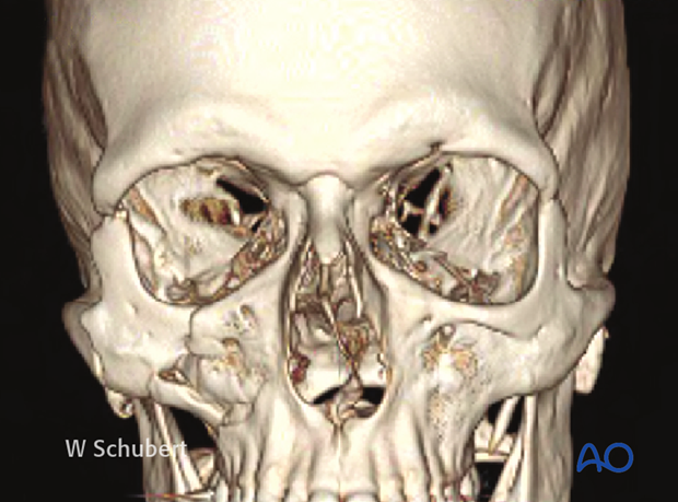
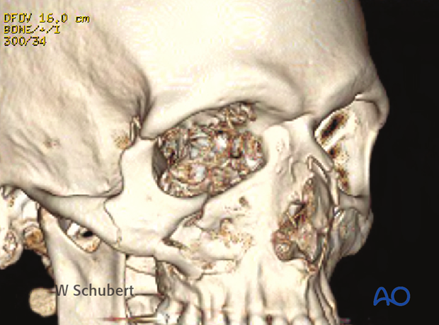
For zygomatic complex and orbital floor fractures, preoperative CT scans in axial and coronal slices are standard. Additional sagittal or oblique parasagittal slices are often very helpful in the assessment of the orbital roof and orbital floor. 3-D reconstructions are also helpful to understand the pattern of displacement and/or rotation.
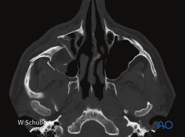
The CT represents an axial slice, and shows a posterior displacement of the zygoma.
This view also shows a fracture through the zygomatic arch.
The coronal slice shows most details of internal orbital disruption and rotation of the zygomatic complex in the coronal plane.
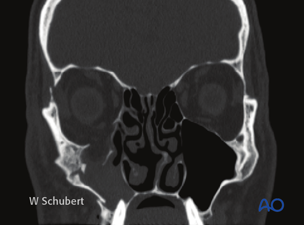
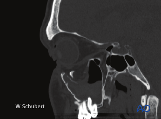
The oblique parasagittal slice is singularly the best view to assess orbital floor fractures. It is also provides an excellent postoperative assessment of the proper placement of an orbital floor plate.
The extent of the orbital floor defect is often underestimated by examination of the preoperative views where the zygoma has been posteriorly displaced. Repositioning of the zygoma anteriorly to its proper location often results in a large orbital floor defect.
Further examinations
The patient needs a complete eye examination because of the nature of the periorbital trauma. It is especially important to make sure that the patient’s visual acuity has not been compromised, and that there is no entrapment of the extraocular muscles (EOM). If there is suspicion of entrapment a forced duction test should be considered.
A fracture of the zygomaticomaxillary complex may commonly cause numbness of the infraorbital nerve distribution.


