Zygoma, zygomatic complex fracture
Definition
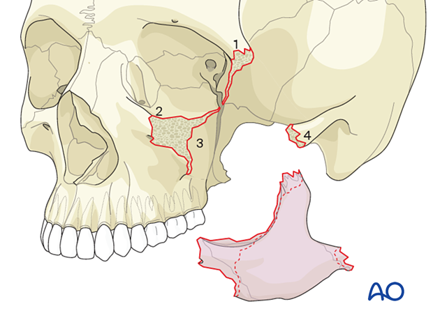
A zygoma fracture is a fracture that separates the zygoma from its surrounding bones. The typical fracture lines of a complete zygoma fracture include the zygomaticofrontal suture, the inferior orbital rim, the zygomaticomaxillary buttress, the zygomatic arch, the orbital floor, and the lateral wall of the orbit at the greater wing of the sphenoid. Internally in the orbit the fracture lines also cross the inferior orbital fissure.
Radiographic findings
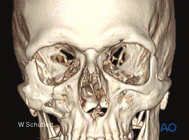
3D reconstruction of a right zygoma fracture.
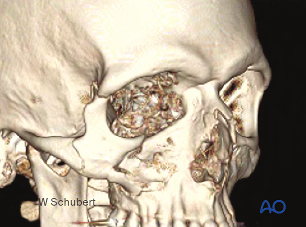
3D reconstruction in a ¾ view of a right zygoma fracture. Note the rotation of the zygoma and the fracture in the inferior orbital rim.
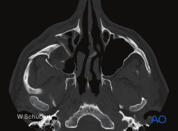
For zygoma and orbital floor fractures, preoperative CT scans in axial and coronal slices are standard. Additional sagittal or oblique parasagittal slices are often very helpful in assessing the orbital roof and orbital floor. 3D reconstructions are also beneficial to understanding the pattern of displacement and rotation.
This CT represents an axial slice and shows a posterior displacement of the zygoma.
This view also shows a fracture through the zygomatic arch.
The coronal slice shows most details of internal orbital disruption and rotation of the zygoma in the coronal plane.
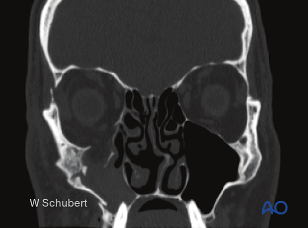
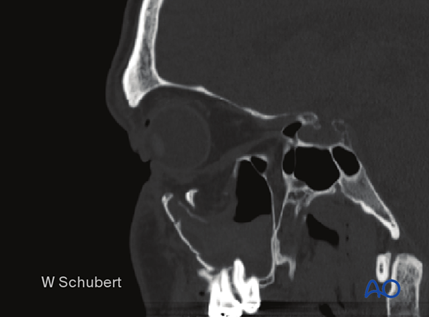
The oblique parasagittal slice is the optimal view for assessing orbital floor fractures. It also helps to provide an excellent postoperative assessment of the proper orbital floor reconstruction.
Examining the preoperative views where the zygoma has been medially and posteriorly displaced often leads to underestimation of the extent of the orbital floor defect. Repositioning of the zygoma anteriorly and laterally into its proper anatomical position results in the creation of a larger orbital floor defect.
Further examination
The patient needs a complete examination of the eye and its adnexa because of the nature of the periorbital trauma. It is crucial to ensure that the patient’s visual acuity has not been compromised and that there is no entrapment of the extraocular muscles (EOM). If there is suspicion of entrapment, a forced duction test will confirm the absence of complete and free extraocular motion.
A fracture of the zygoma commonly causes numbness in the distribution of the infraorbital nerve.


