Palatoalveolar fracture, simple
Definition
A simple palatoalveolar fracture divides the palate into two palatoalveolar fragments.
Simple (noncomminuted) palatal fractures are commonly associated with Le Fort I fractures but can be seen with other midface complex fractures. They are almost always longitudinal, and in adults, parallel to the center. In children, where the midline palatal suture is open, they divide at the center. Rarely, palatal fractures are transverse or oblique.
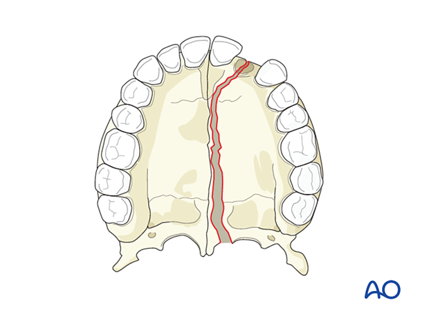
The patients typically complain of malocclusion. There is almost always a laceration over the fracture in the palate, and frequently a laceration of the upper lip. Generally, one side is displaced more than the other.
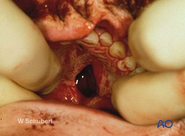
In the case of significant splaying of the palatal fracture, there is often, as stated above, a tear through the palate's mucosa, which can be observed on physical examination.
Diagnosis may be confirmed with CT imaging. Ideal views include the axial and coronal cuts.
This clinical photograph shows the tear in palatal mucosa.
Radiographic findings
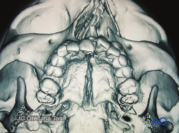
3D CT scan
The 3D reconstruction shows the palatal fracture pattern and displacement separated parallel to the midline suture.
2D CT scans
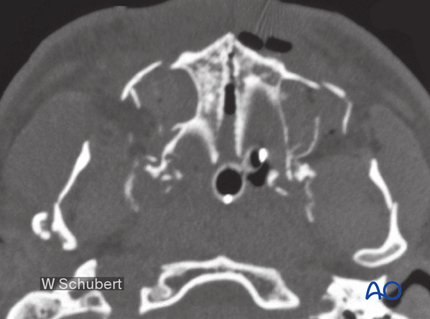
Preoperative axial CT scan.
This CT shows significant splaying of the palate in the midline. It also shows a Le Fort I fracture and significant mandibular fractures.
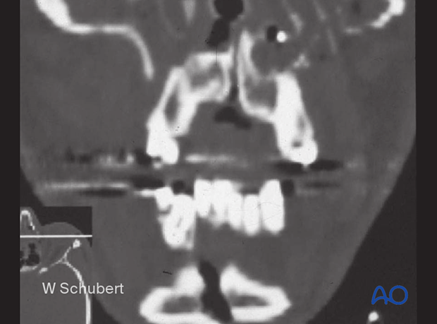
Coronal view of the same case. This fracture is just off the midline.
This CT shows splaying of the palate in the midline and a mandibular symphyseal fracture with similar splaying.
3D CT scan
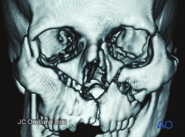
In most cases, this fracture is associated with other midface fractures.
In this case, we find a right sided Le Fort III (zygomatic fracture) associated with Le Fort II and I fractures and an NOE fracture on the left side.
