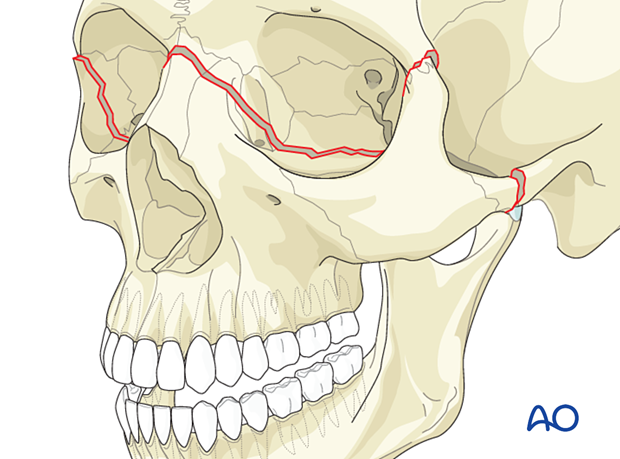
Le Fort III fracture
Definition
The Le Fort III fracture is also referred to as craniofacial disjunction. The fracture line begins at the frontozygomatic suture traversing the lateral aspect of the internal orbit, continuing along the sphenozygomatic suture to the inferior orbital fissure, extending medially across the floor of the orbit, then up the medial wall of the orbit across the dorsum of the nose and proceeding to the contralateral side of the face. Because injury forces are usually laterally oriented, it is common to see a Le Fort III level fracture on one side of the face and a Le Fort II level fracture on the other. Generally, Le Fort III fractures are not single segment, but are comminuted, with Le Fort II and Le Fort I segments as lesser individual components of the fracture. Special attention must be paid to nasal or pharyngeal airway obstruction caused by bleeding.
Patients with Le Fort III injuries are often admitted to the hospital unconscious and intubated. Foreign bodies such as teeth or tooth fragments may obstruct the airway.
Severe bleeding and CSF leakage may accompany Le Fort II and III fractures and affect the treatment and outcome.
It should be recognized that Le Fort III fractures involve the orbit. Basic assessment of visual acuity is mandatory in the conscious patient. In the unconscious patient, the swinging-flashlight test can give evidence of or exclude afferent pupillary defects (Marcus Gunn pupil).
It should be emphasized that all of the Le Fort fractures go through the pterygoid plates.
When assessing Le Fort fractures, it is important to ascertain the patient’s premorbid occlusion.
CSF leak
The following diagnostic procedures can be performed if there is a suspected CSF leak (clinical sign: straw-colored nasal drainage):
- Application of fluorescent dyes
- Comparison of the concentration of glucose between fluid and patient’s serum
- Laboratory analysis for beta-2 transferrin
- Specialized neuroradiological imaging with intrathecal contrast medium
Radiographic findings
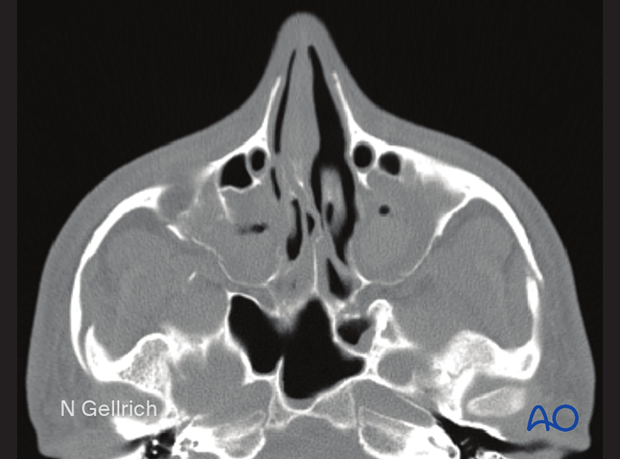
CT slices showing the typical fracture pattern of a Le Fort III fracture.
Note the fracture in the lateral wall of the orbit which extends to the frontozygomatic suture.
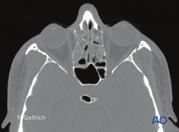
Note the fracture of the zygomatic arch near the temporal bone on the right side. Likewise, on the left side, note the fracture of the zygomatic arch.
3D CT scan
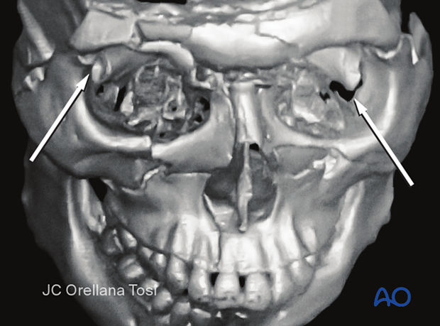
Note the fracture of the frontozygomatic articulation and how the fracture goes through the floor of the orbit, continues through the medial wall, and separates the radix of the nose through the other side, making it a complete disjunction of the face from the skull. In this example, the superior orbital rim, skull base, and frontal sinus are also fractured.
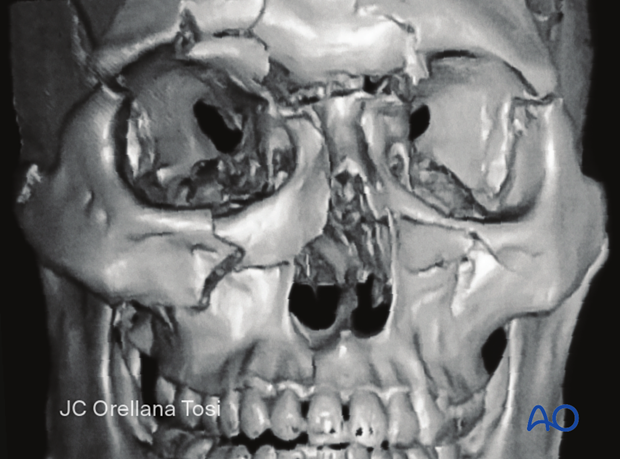
Separation of the radix of the nose from the frontal bone.
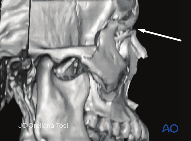
This image shows the fracture spreading through the orbit.
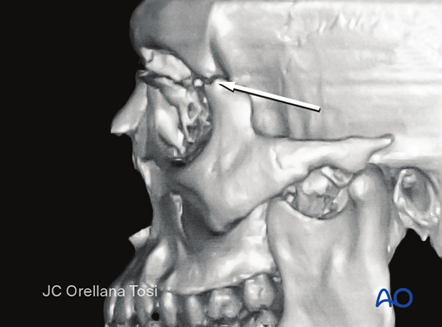
Note how the fracture goes through both orbits and separates the malar bones from the skull.
