Le Fort I fracture, unilateral comminution
Definition
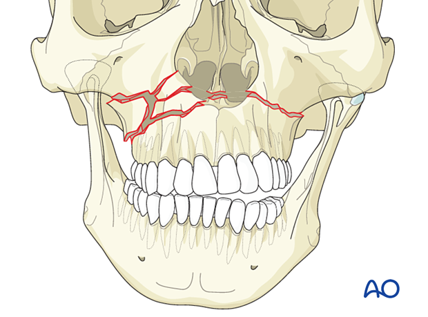
A comminuted Lefort I fracture extends from the piriform aperture through the medial and lateral maxilla and the lateral nasal walls. It extends posteriorly to involve the pterygoid region on both sides. The example shows a right comminuted fracture and a simple left Le Fort I fracture.
These fractures can occur without bone defects, in which case the fragments can be fixed to the plates. If there are bone defects the fragments are not available for fracture fixation or bone buttressing; grafts are therefore necessary to restore the vertical buttresses of the midface.
This illustration shows a Le Fort I fracture with unilateral comminution (without bone defect).

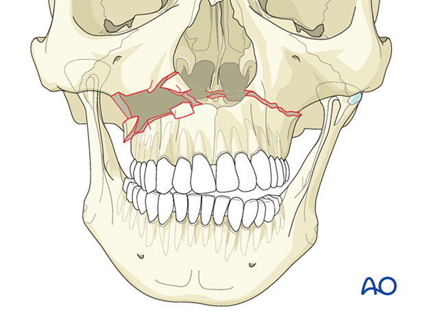
In cases of comminution with excessively small fragments or missing bone, surgical reconstruction may require a bone graft to replace or further stabilize small fragments. If the small fragments are removed in the exposure, a defect fracture is created which requires replacement of the fragments and/or graft at the Le Fort I level.
Cautious and gentle exposure at the Le Fort I level allows maximal preservation of small bone segments, including their orientation, and may minimize fracture defects and the need for bone grafts.
This illustration shows a Le Fort I fracture with unilateral comminution with a defect. There is also premature occlusal contact on the defect side and an open bite on the non-defect side.

Radiographic findings
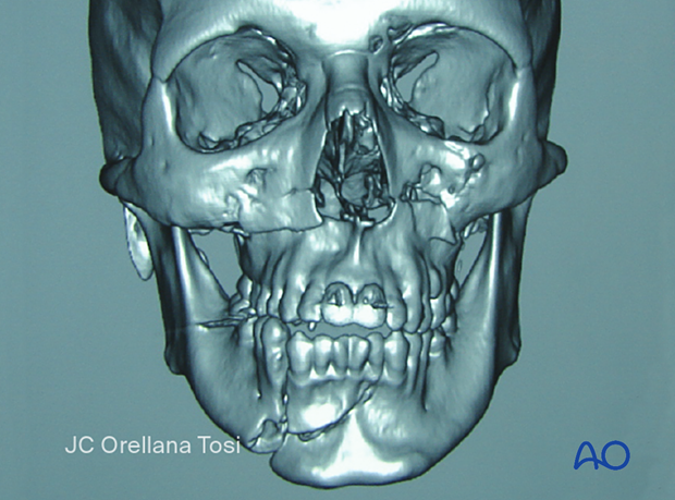
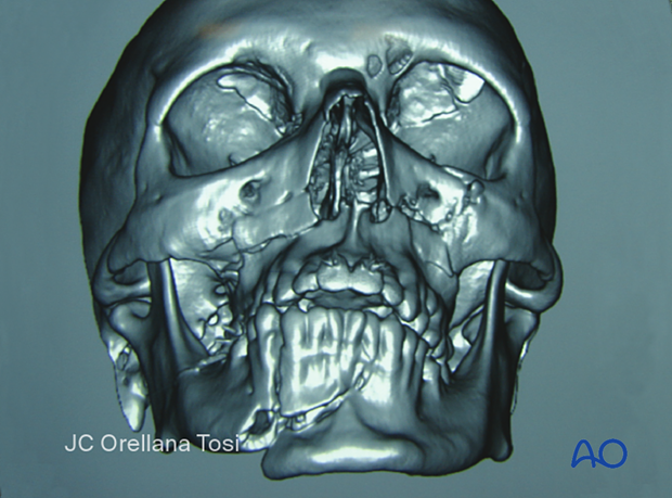
A 3D CT scan shows a Le Fort I fracture in a frontal view. There is comminution on the right side. The maxillary fracture is also associated with multiple mandibular fractures.

In this image, a multifragmentary (comminuted) Le Fort I fracture is visualized.


